Courtesy: Amr Abdelgawad, Maimonaides Medical Centre, Brooklyn, NY, USA
Cavus Foot (High-Arched Foot)
Introduction
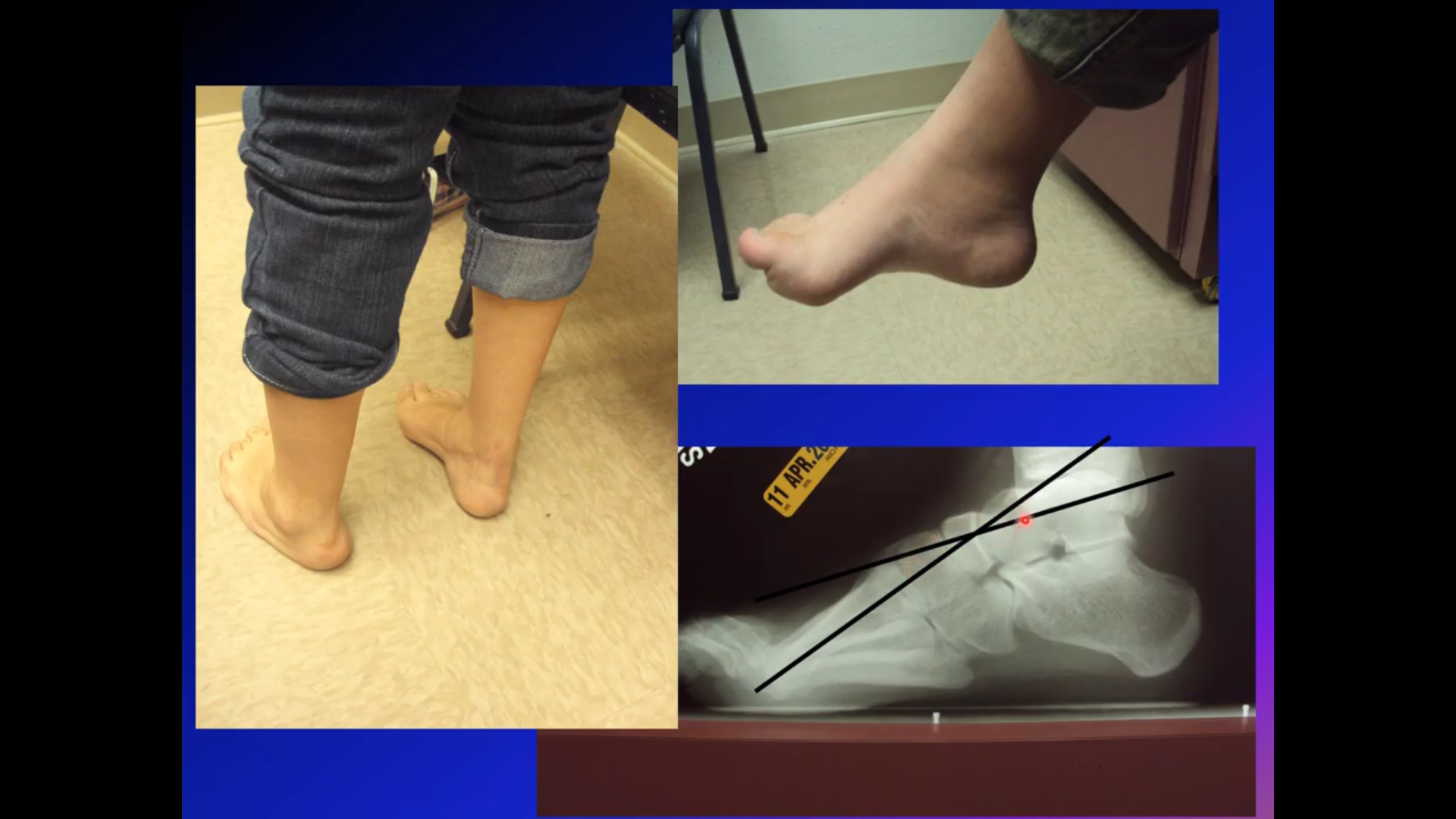
Cavus foot refers to an abnormally high medial longitudinal arch of the foot.
It is essentially the opposite of flatfoot deformity and is commonly associated with underlying neurological disorders.
In many patients, cavus foot may be the first or only sign of a neurological condition.
Etiology
Neurological Causes
Neurological disorders are the most common causes of cavus foot.
Important causes include:
- Charcot-Marie-Tooth disease
- Tethered cord syndrome
- Diastematomyelia
- Poliomyelitis
- Spinal cord pathology
Important Clinical Principle
Cavus foot should always prompt careful neurological evaluation, especially in:
- Progressive deformity
- Unilateral deformity
- Newly developing cavus foot
Pathophysiology
Muscle Imbalance
The deformity develops due to imbalance between muscle groups.
A classic imbalance is:
- Peroneus longus overpowering tibialis anterior
Resulting Deformity
This imbalance produces:
- Plantar flexion of the first ray
- Hindfoot varus
- Cavovarus deformity
Over time, the deformity may become rigid and progressive.
Cavovarus Foot
Most cavus feet are actually:
- Cavovarus feet
meaning there is both:
- High arch
- Hindfoot varus
Tripod Theory of the Foot
Normal Foot Mechanics
Normally, body weight is distributed across three points:
- Heel
- First metatarsal head
- Fifth metatarsal head
This is known as the tripod configuration.
Effect of Cavus Foot
In cavus deformity:
- Weight distribution becomes abnormal
- Forefoot overload occurs
- Lateral border overload may develop
This contributes to instability and pain.
Clinical Features
Common Findings
Patients may present with:
- High medial arch
- Cavovarus alignment
- Claw toes
- Callosities
- Lateral foot pain
- Ankle instability
Neurological Assessment
A complete neurological examination is essential to assess for:
- Muscle weakness
- Sensory loss
- Reflex abnormalities
- Spinal pathology
Coleman Block Test
Purpose
The Coleman block test evaluates flexibility of the hindfoot varus deformity.
Technique
The patient stands with:
- Heel and lateral border of foot supported
- First metatarsal allowed to hang free
Interpretation
Varus Corrects
If hindfoot varus corrects:
- Deformity is flexible
- Forefoot-driven cavus deformity is likely
Varus Persists
If hindfoot varus remains:
- Hindfoot deformity is rigid
This influences surgical planning.
Radiographic Evaluation
Meary’s Angle
Meary’s angle is formed between:
- Long axis of talus
- First metatarsal axis
Normal Value
Normal Meary’s angle is approximately:
- 0–5°
Abnormal Findings
An increased angle indicates:
- Cavus deformity
Radiographs also help assess:
- Hindfoot alignment
- Forefoot plantar flexion
- Degenerative changes
Management Principles
Step 1 – Identify the Underlying Cause
Because many cavus feet are neurological in origin:
- Neurological evaluation is essential
This may include:
- Neurology consultation
- Spine imaging
- Electrophysiological studies
Conservative Treatment
Bracing and Orthotics
Non-operative treatment may help in flexible deformities.
Common options include:
- Lateral heel wedge
- Custom orthotics
- Bracing
Important Orthotic Principle
Support under the first metatarsal head is generally avoided because:
- It may worsen the cavus deformity
Surgical Treatment
Progressive Nature
Most neurological cavus deformities are progressive and may eventually require surgery.
Surgical Goals
Surgery aims to:
- Correct deformity
- Balance muscle forces
- Improve function
- Prevent recurrence
Common Surgical Procedures
Plantar Fascia Release
Useful for:
- Flexible cavus deformity
- Soft tissue tightness
First Metatarsal or Cuneiform Osteotomy
Performed to correct:
- Plantar-flexed first ray
Calcaneal Osteotomy
Indicated for:
- Fixed hindfoot varus
Helps restore hindfoot alignment.
Tendon Transfers
Used to:
- Rebalance muscle forces
- Reduce progression
- Improve function
Prognosis
Outcome depends on:
- Severity of deformity
- Flexibility
- Underlying neurological condition
- Timing of intervention
Progressive neurological disorders may continue to influence long-term results.
Key Clinical Pearls
- Cavus foot is commonly neurological in origin.
- Cavovarus is the most frequent deformity pattern.
- Always perform a neurological examination.
- Coleman block test determines hindfoot flexibility.
- Meary’s angle helps quantify cavus deformity.
- Early orthotic management may help flexible deformities.
- Many progressive deformities ultimately require surgery.
Final Take-Home Message
Cavus foot is a high-arched deformity most commonly caused by neurological disease.
The deformity results from muscle imbalance and frequently progresses over time.
Accurate evaluation should include:
- Neurological assessment
- Clinical examination
- Flexibility testing
- Radiographic analysis
Treatment ranges from orthotics and bracing to complex reconstructive surgery depending on severity and underlying cause.
Related Posts
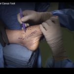 Triple Arthrodesis for Cavus Foot
Courtesy: Anand Vora, MD, (Chicago, IL) and Arthrex Inc
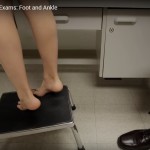 Paediatric Foot and Ankle Exam
Courtesy: David Horn, MD, orthopedic surgeon at The Children's Hospital of Philadelphia http://chop.edu/ortho
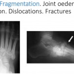 Charcot's Foot
Charcot foot from Shewei Aziz Courtesy: Dr Sheweidin Aziz, Registrar in Orthopaedics and Trauma, Lincolnshire,…
Leave a Reply