GLENOHUMERAL ARTHRITIS Aetiology: Osteoarthritis(associated with posterior glenoid wear) Rheumatoid arthritis( assoc. with central glenoid wear) Secondary degenerative joint disease -Repetitive and major trauma -End-stage AVN -Rotator cuff tear arthropathy -Arthritis associated with instability or surgery for instability Clinical Features: – Pain and stiffness in the shoulder – adduction and internal rotation deformity of the joint […]
Evidence Based Orthopaedic Principles
Ultrasound Therapy
ULTRASONIC THERAPY Ultrasound is an electromagnetic wave with frequencies above 20,000Hz For therapy the frequency of sound waves used are between 85kHz to 3MHz and delivered at intensities between 0 and 3W/sq.cm Production: When electrical energy is applied on piezoelectric crystals they produce vibration and mechanical deformity of their molecular structure. This phenomenon is […]
Tibial Plateau Fractures
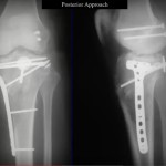
Tibial Plateau Fracture Anatomical Pearls: The articular surface of the lateral tibial plateau is flat or slightly convex in relation to the medial tibial plateau that is concave, which provides greater congruity with the medial femoral condyle than on the lateral side. The lateral plateau is also higher than the medial plateau, accounting for the […]
Stem Cells in Orthopaedic Surgery
Stem Cells in Orthopaedic Surgery A stem cell is an ‘immature’ or undifferentiated cell which is capable of producing an identical daughter cell. Stem cells must have a capacity for self-renewal giving rise to more stem cells, and the ability to differentiate into tissues of various lineages under appropriate condition They may be totipotent, pluripotent […]
Lisfranc Joint Injury
The Lisfranc joint complex has a relatively rigid medial column(1st, 2nd and 3rd tarsometatarsal joints) and a mobile lateral column…..
Unicameral Bone Cyst
Unicameral bone cysts are developmental anomalies of the physis where there is a transient failure of ossification of physeal cartilage and cyst formation…..
Piriformis Syndrome
Yeoman first described the relationship between the sciatic nerve and piriformis muscle and Robinson coined the term ‘piriformis syndrome’…..
Slipped Capital Femoral Epiphysis(SCFE)
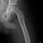
Slipped capital femoral epiphysis refers to the atraumatic separation of the epiphysis in the epiphyseal plate of the femoral neck with displacement of the femoral head, usually in a medial and dorsal direction, during the pubertal growth spurt……
Congenital Muscular Torticollis
Congenital muscular torticollis or congenital wry neck, is the most common cause of torticollis in the infant and young child, presenting at a median age of 2 months
Rotator cuff Tears
The primary mechanical function of the rotator cuff is to balance the force couples about the glenohumeral joint to provide a stable fulcrum of motion and functional glenohumeral kinematics…..
POSITRON EMISSION TOMOGRAPHY(PET Scan)
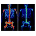
POSITRON EMISSION TOMOGRAPHY PET allows the visualisation of the metabolic activity of disease. Helps in diagnosis of malignant tumours and their recurrence. Also helps in the staging of tumours and the monitoring of their response to therapy, and in the diagnosis of osteomyelitis. The positron-emitting tracers may be used to define: Amino-acid uptake (11C-methionine; […]
MADELUNG’S DEFORMITY
CONGENITAL SUBLUXATION OF THE WRIST (MADELUNG’S DEFORMITY) Pathogenesis (Brailsford): Stunted development of inner third of the growth cartilage at the lower end of the radius, due to still unknown cause. Growth of the outer two-thirds continues and, as a result, the radial shaft is bowed backwards, the interosseous space is increased, there is overgrowth of […]
Fibrous Dysplasia
Fibrous Dysplasia originally described by Lichtenstein in 1938 and by Lichtenstein and Jaffe in 1942 represent approximately 5% to 7% of benign bone tumors A sporadic disorder of osseous and fibrous tissue development characterised by postzygotic mutation of GNAS1 gene coding for stimulatory G protein. Analysis of the Gs ? subunit in patients with fibrous […]
Intercondylar Elbow Fractures
INTERCONDYLAR FRACTURES OF THE ELBOW Mechanism of injury: Is by a force directed towards an elbow which is flexed > 90° which causes the ulna to drive against the trochlea Riseborough and Radin Classification • Type I: Nondisplaced • Type II: Slight displacement with no rotation between the condylar fragment. • Type III: Displacement with […]
Calcaneal Fractures
© Hitesh Gopalan U Excerpts from “Orthopaedic Principles- A Review” CALCANEUM fractures Mechanism of Injury: fall from height or motor vehicle accident Classification: a. Extra Articular Fractures • Anterior process fractures • Tuberosity fractures • Medial process fractures • Sustentacular fractures • Body fractures not involving the subtalar articulation b. Intra Articular Fractures (Essex Lopresti […]