Courtesy: Prof Nabile Ebraheim, University of Toledo, Ohio, USA STERNOCLEIDOMASTOID MUSCLE ANATOMY The sternocleidomastoid muscle (also known as sternomastoid) is one of the largest and most superficial cervical muscles located in the superficial layers on the side of the neck. ORIGIN The sternocleidomastoid muscle arises from the medial portion of the clavicle and the […]
-Applied Anatomy
Anatomy of Flexor Digitorum Profundus
ANATOMY OF FLEXOR DIGITORUM PROFUNDUS The flexor digitorum profundus (FDP) is a muscle in the forearm that flexes the fingers. The flexor digitorum profundus muscle lies in the deep compartment of the forearm. ORIGIN The FDP muscle arises from the upper 3/4 of the volar and medial surfaces of the ulna as well as […]
Anatomy of the #Soleus Muscle
Courtesy: Prof Nabile Ebraheim, University of Toledo, Ohio, USA GENERAL ANATOMY The soleus is a broad, flat muscle located beneath the gastrocnemius muscle. It lies within the superficial posterior compartment of the leg. The soleus is a major contributor to the triceps surae muscle complex. COMPARTMENTS OF THE LEG The leg is divided into four […]
Anatomy of the Teres Major
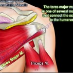
Anatomy of the Teres Major Muscle Overview The teres major is a thick, powerful muscle of the posterior axillary region. It is one of the muscles connecting the scapula to the humerus and plays an important role in movements of the shoulder joint. Origin and Insertion Origin:Dorsal (posterior) surface of the inferior angle of […]
Anatomy of the Sartorius Muscle
Courtesy: Prof Nabile Ebraheim, University of Toledo, Ohio, USA Origin- Sartorius muscle arises from the anterior superior iliac spine of the pelvic bone. The sartorius muscle crosses the upper third of the thigh obliquely, downwards medially and then descends vertically towards its insertion. It is a superficial muscle, the longest muscle and it’s fibres are […]
Anatomy of the Psoas and Iliacus
Courtesy: Prof Nabile Ebraheim, University of Toledo, Ohio, USA
Adductor Magnus Anatomy
Courtesy: Prof Nabile Ebraheim, University of Toledo, Ohio, USA
Posterior Triangle of the Neck- #Anatomy
Anatomy of #Gluteus Maximus
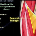
Anatomy of the #Femoral Triangle
Courtesy: Prof Nabile Ebraheim, University of Toledo, Ohio, USA
Joint Biomechanics
Joint biomechanics from Lennard Funk Courtesy: Prof Lennard Funk, Wrightington Upper Limb Unit, Wrightington, UK
Structures passing through Greater Sciatic Notch
Courtesy : Prof Nabile Ebraheim, University of Toledo, Ohio, USA
Musculoskeletal Histology: Clinical Correlation
By Dr Aaron Rosenberg,MD Professor, University of Miami, Jackson Memorial Hospital, Florida,USA Courtesy : Dr Sanjay Chaturvedi, Organising Secretary, IOACON 2013, Agra
Hip Joint Movements
Courtesy : Prof Nabile Ebraheim, University of Toledo, Ohio, USA
Obturator Internus Anatomy
Obturator internus is a fan shaped muscle that passes through lesser sciatic notch. The muscle lies within the pelvis and the tendon lies posterior to the hip joint. Origin and insertion: The obturator internus arises from the medial surface of the obturator membrane, ischium, and the rim of pubis. The obturator internus muscle ends […]