Courtesy: Rishi Dhir, FRCS Orth, Consultant Upper Limb Surgeon, Princess Alexandra Hospital, Harlow, UK
Genetics
Chromosome Structure
- Chromosomes are structures in the nucleus containing genetic material
- Components include:
- DNA
- RNA
- Histone proteins
- Non-histone proteins
Human Karyotype
- Normal somatic cell contains 46 chromosomes
- 22 pairs of autosomes
- 1 pair of sex chromosomes
- Sex chromosomes:
- XX ? Female
- XY ? Male
Chromosome Anatomy
- Chromosome has:
- Short arm (p arm)
- Long arm (q arm)
- Arms separated by centromere
Chromosome Types (based on centromere location)
- Metacentric
- Centromere near middle
- p and q arms equal length
- Submetacentric
- Arms unequal
- Acrocentric
- Centromere near one end
- Very short p arm
- Telocentric
- Centromere at terminal end
DNA Structure
DNA Composition
DNA consists of:
- Nitrogenous base
- Sugar (deoxyribose)
- Phosphate backbone
Nitrogenous Bases
Purines
- Adenine (A)
- Guanine (G)
Pyrimidines
- Cytosine (C)
- Thymine (T)
Base Pairing
- Adenine pairs with Thymine
- Guanine pairs with Cytosine
Structure
- DNA forms a double helix
- Bases linked by hydrogen bonds
Cell Division
Mitosis
- Produces two identical daughter cells
- DNA replication occurs before division
- Replication is semi-conservative
- Each daughter strand contains one original strand
Steps:
- DNA replication
- Chromosomes align in cell center
- Chromatids separate
- Two identical cells formed
RNA and Protein Synthesis
Transcription
- DNA ? mRNA
- Enzyme: RNA polymerase
Translation
- mRNA moves to cytoplasm
- Combines with:
- Ribosomal RNA
- Transfer RNA
- Produces proteins
Key Definitions
- Replication: DNA ? DNA (DNA polymerase)
- Transcription: DNA ? RNA
- Translation: RNA ? Protein
Chromosomal Abnormalities
Numerical Abnormalities (Aneuploidy)
Monosomy
- Loss of one chromosome
- Example:
- Turner syndrome (XO)
Features of Turner syndrome:
- Short stature
- Webbed neck
- Cubitus valgus
- Coarctation of aorta
- Broad chest with widely spaced nipples
- Amenorrhea and infertility
Trisomy
- Gain of one chromosome
Examples:
Down syndrome (Trisomy 21)
- Intellectual disability
- Characteristic facial features
- Congenital heart disease
- Wide gap between first and second toes
Klinefelter syndrome (XXY)
- Male with extra X chromosome
- Features:
- Gynecomastia
- Female body habitus
- Sparse body hair
- Testicular atrophy
- Infertility
Structural Chromosome Abnormalities
Definitions
Genotype
- Genetic constitution of an individual
Phenotype
- Clinical expression of genotype
Allele
- Alternative form of a gene at same locus
Homozygous
- Two identical alleles
Heterozygous
- Two different alleles
Mutation Types
Point mutation
- Single nucleotide change
Deletion
- Loss of chromosome segment
Inversion
- Segment rotated 180°
Translocation
- Genetic material transferred between chromosomes
Modes of Inheritance
Meiosis
- Specialized cell division producing gametes
- Results in haploid cells (23 chromosomes)
Key Features
- Crossing over occurs at chiasma
- Produces genetic variation
Sources of variation:
- Crossing over
- Independent segregation
Mendelian Inheritance
Autosomal Dominant
Characteristics:
- Only one abnormal allele required
- Affected parent ? 50% chance of affected child
- Male : Female ratio 1:1
Examples:
- Achondroplasia
- Marfan syndrome
- Neurofibromatosis type 1
- Osteogenesis imperfecta
Autosomal Recessive
Characteristics:
- Two abnormal alleles required
- Parents usually carriers
If both parents are carriers:
- 25% affected
- 50% carriers
- 25% normal
Example:
- Sickle cell disease
X-Linked Recessive
- More common in males
- Males have only one X chromosome
Examples:
- Hemophilia
- Duchenne muscular dystrophy
Inheritance patterns:
Mother carrier + father normal
- 50% sons affected
- 50% daughters carriers
- No daughters affected
Father affected + mother normal
- All daughters carriers
- All sons unaffected
X-Linked Dominant
Example:
- Hypophosphatemic rickets
Inheritance:
Mother affected:
- 50% sons affected
- 50% daughters affected
Father affected:
- All daughters affected
- No sons affected
Penetrance and Expressivity
Penetrance
- Probability that genotype will express phenotype
Incomplete penetrance
- Gene present but no clinical expression
Variable Expressivity
- Same genotype
- Different severity of symptoms
Example:
- Marfan syndrome
Polygenic Inheritance
- Multiple genes influence phenotype
- Environmental factors may contribute
Examples:
- Developmental dysplasia of hip (DDH)
- Clubfoot (Talipes equinovarus)
Key feature:
- Risk decreases in subsequent generations
Embryology of the Limb
Limb Bud Development
- Occurs 4–8 weeks gestation
Rule of Threes
- Three zones
- Three genes
- Three directions
Zones
Apical Ectodermal Ridge (AER)
- Controls limb growth
Progress Zone
- Cells undergo proliferation
Zone of Polarizing Activity (ZPA)
- Controls anterior-posterior development
Growth Directions
Proximal ? Distal
Controlled by:
- HOX (homeobox) genes
Posterior ? Anterior
Controlled by:
- Sonic hedgehog gene
Associated abnormalities:
- Radial club hand
- Thumb hypoplasia
- Fibular hemimelia
Dorsal ? Ventral
Controlled by:
- WNT (wingless type) gene
Limb Bud Associations
Congenital anomalies often associated with VACTERL association
VACTERL stands for:
- Vertebral anomalies
- Anal atresia
- Cardiac defects
- Tracheoesophageal fistula
- Renal anomalies
- Limb abnormalities
Important exam principle:
- Associated anomalies may be life-threatening, not limb deformity.
Embryology of the Spine
Gastrulation
Occurs during week 3
Three germ layers form:
- Ectoderm
- Mesoderm
- Endoderm
Germ Layer Derivatives
Ectoderm
- Nervous system
- Skin epidermis
Mesoderm
- Muscle
- Bone
- Connective tissue
Endoderm
- Gastrointestinal lining
- Respiratory tract lining
Vertebral Development
Key structures:
- Notochord
- Somites
Notochord
- Releases sonic hedgehog signals
- Forms nucleus pulposus
Somites
Differentiate into:
- Dermatome ? Skin
- Myotome ? Muscle
- Sclerotome ? Vertebrae and annulus fibrosus
Neural Tube Development
Process called neurulation
Steps:
- Neural plate formation
- Neural folds develop
- Fusion ? neural tube
Neural Tube
Forms:
- Brain
- Spinal cord
Neural Crest
Forms:
- Peripheral nervous system
Spina Bifida
Failure of neural tube closure
Types:
- Spina bifida occulta
- Meningocele
- Myelomeningocele
Clinical sign:
- Hair tuft over spine
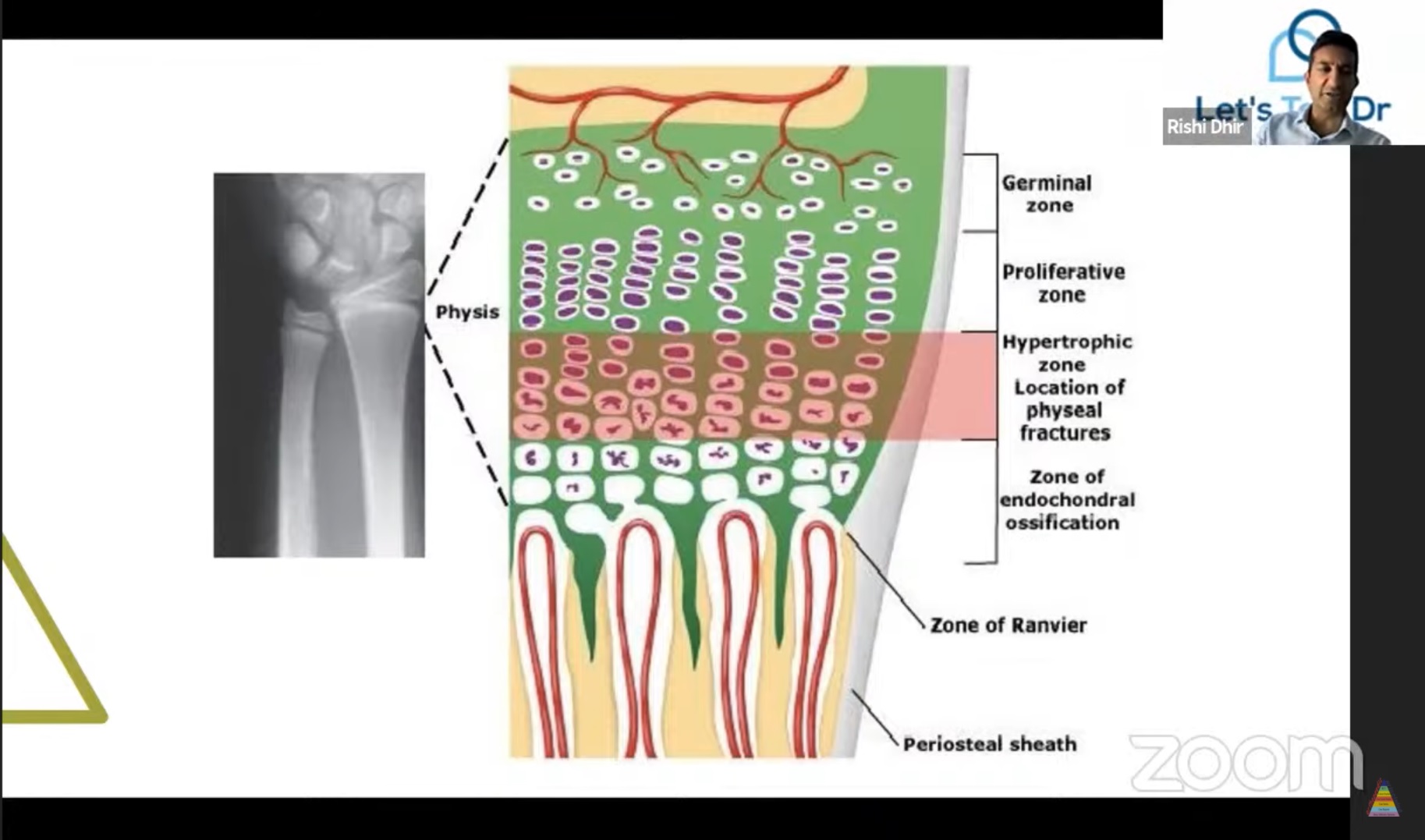
Physis (Growth Plate)
Growth Plate Zones
Resting (Germinal) Zone
- Storage zone
- Contains glycogen, lipids
Associated disorders:
- Storage diseases
- Pseudoachondroplasia
- Diastrophic dysplasia
Proliferative Zone
- Chondrocyte multiplication
Associated disorders:
- Gigantism
- Achondroplasia
Hypertrophic Zone
- Cells enlarge
- Site of physeal fractures
Subdivisions:
- Zone of maturation
- Zone of degeneration
- Zone of provisional calcification
Metaphysis
Contains:
- Primary spongiosa
- Secondary spongiosa
Common site of:
- Osteomyelitis
Reason:
- Hairpin vascular loops
- Slow blood flow
- Bacterial deposition
Salter–Harris Fractures
Salter–Harris II
- Through physis + metaphysis
- Germinal layer preserved
- Better prognosis
Salter–Harris III
- Through physis + epiphysis
- Involves germinal layer
- Worse prognosis
Complications of Physeal Injuries
Complete Physeal Arrest
- Limb shortening
Partial Physeal Arrest
- Angular deformity
Management of Physeal Bar
Evaluation:
- MRI
- CT scan
Treatment depends on:
- Patient age
- Size of physeal bar
- Remaining growth
Options:
- Bar excision + interposition graft
- Completion epiphysiodesis
- Contralateral epiphysiodesis
- Corrective osteotomy
Leave a Reply