• Skip to main content
  • Skip to secondary menu
  • Skip to primary sidebar
OrthopaedicPrinciples.com

OrthopaedicPrinciples.com

Integrating Principles and Evidence

Integrating Principles and Evidence

  • Home
  • Editorial Board
  • Our Books
    • Evidence Based Orthopaedic Principles
  • Courses
  • Exams
  • Reviews
  • Live Program
  • Contact

Anatomy Of The Teres Minor Muscle

Courtesy: Prof Nabil Ebraheim, University of Toledo, Ohio, USA

Anatomy of the teres minor muscle

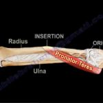
  • The teres minor is a narrow muscle which originates from the posterior lateral surface of the
    scapula and inserts into the greater tuberosity of the humerus .
  • Teres minor is innervated by the posterior branch of axillary nerve .
  • The same nerve that gives cutaneous innervation to the lateral part of the shoulder .

Important anatomical spaces

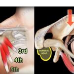
  • Teres minor contribute to important anatomical spaces in the posterior part of the shoulder such as the quadrangular space
  • Boundaries of quadrangular space : teres minor superiorly , teres major inferiorly , long head of triceps medially , surgical neck of humerus laterally.
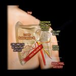
  • The quadrangular space contains the posterior humeral circumflex artery and axillary nerve . The
    tere minor muscle also contributes to the triangular space .
  • Boundaries of triangular space : teres major , teres minor and long head of triceps . This triangular
    space also contains circumflex scapular artery . The teres minor muscle does not contribute to the
    triangular interval .

Boundaries of triangular interval : teres major , humeral shaft , long head of triceps .

  • The triangular interval contains the radial nerve and deep branch of brachial artery
  • The teres major muscle contributes to 3 important anatomical spaces while the teres minor only
    contributes to 2 important anatomical spaces.
  • The good way to see the teres minor muscle is MRI sagittal view .The infraspinatous is the main
    external rotator of the shoulder with the arm to the side(adducted).The teres minor muscle is the
    main external rotator of the shoulder with the arm abducted.

Clinical examination

  • Horn Blowers test-The test is used to determine the strength of the teres minor muscle. The patient
    and the examiner are both standing and the patients arm is elevated to 90 degrees,the patients
    elbow is then flexed to 90 degrees and the patient is asked to laterally rotate the shoulder.
    Weaknesses and /pain against resistance signals a positive test.

Why teres minor is important?

  • Innervation of the teres minor muscle is unexpected because it comes from the posterior branch of
    axillary nerve .
  • It is important in posterior approach to the shoulder joint, yhe approach is usually
    done in the interval between the infraspinatous and teres minor muscle.
  • If you go below the teres minor muscle then you could be in the quadrangular space and could injure the neurovascular
    structure including the axillary nerve.
  • Check if the teres minor muscle is atrophic on the MRI,atrophy of the muscle can be idiopathic or it may be due to quadrangular space impingement or syndrome.
  • Idiopathic atrophy is associated with rotator cuff pathology.

Post Views: 4,706

Related Posts

  • Anatomy of the Pronator Teres Muscle

    Courtesy: Prof Nabil Ebraheim, University of Toledo, Ohio, USA   ANATOMY OF PRONATOR TERES MUSCLE…

  • Anatomy Of The Teres Major Muscle

    Courtesy: Prof Nabil Ebraheim, University of Toledo, Ohio, USA Anatomy of Teres Major Muscle Origin-…

  • Anatomy Of The Pectoralis Minor Muscle

    Courtesy: Prof Nabil Ebraheim, University of Toledo, Ohio, USA ANATOMY OF PECTORALIS MINOR Pectoralis minor…

Reader Interactions

Leave a Reply

Your email address will not be published. Required fields are marked *

Primary Sidebar

Follow Us

instagram slideshare

Categories

  • -Applied Anatomy
  • -Approaches
  • -Basic Sciences
  • -Cartilage & Meniscus
  • -Classifications
  • -Examination
  • -Foot and Ankle
  • -Foot and Ankle Trauma
  • -FRCS(Tr and Orth) tutorials
  • -Gait
  • -Hand and Wrist
  • -Hand and Wrist Trauma
  • -Hand Infections
  • -Hip and Knee
  • -Hip Preservation
  • -Infections
  • -Joint Reconstruction
  • -Knee Arthroplasty
  • -Knee Preservation
  • -Metabolic Disorders
  • -Oncology
  • -OrthoBiologics
  • -OrthoPlastic
  • -Paediatric Orthopaedics
  • -Paediatric Trauma
  • -Patellofemoral Joint
  • -Pelvis
  • -Peripheral Nerves
  • -Principles
  • -Principles of Surgery
  • -Radiology
  • -Rheumatology
  • -Shoulder and Elbow
  • -Shoulder and Elbow Arthroplasty
  • -Spine Deformity
  • -Spine Oncology
  • -Spine Trauma
  • -Spine, Pelvis & Neurology
  • -Sports Ankle and Foot
  • -Sports Elbow
  • -Sports Knee
  • -Sports Medicine
  • -Sports Medicine Hip
  • -Sports Shoulder
  • -Sports Wrist
  • -Statistics
  • -Technical Tip
  • -Technology in Orth
  • -Trauma
  • -Trauma (Upper Limb)
  • -Trauma Life Support
  • -Trauma Reconstruction
  • Book Shelf
  • Book Shelf Medical
  • Careers
  • Case Studies and Free Papers
  • DNB Ortho
  • Evidence Based Orthopaedic Principles
  • Evidence Based Orthopaedics
  • Exam Corner
  • Fellowships
  • Guest Editor
  • Guest Reviews
  • Image Quiz
  • Instructional Course Lectures
  • Journal Club
  • MCQs
  • Meetings and Courses
  • Multimedia
  • News and Blog
  • Plaster Techniques
  • Podcasts
  • Public Health
  • Rehabilitation
  • Research
  • Shorts and Reels
Copyright@orthopaedicprinciples.com. All right rerserved.