![]()
Courtesy: Prof Nabile Ebraheim, University of Toledo, Ohio, USA
GENERAL ANATOMY
-
The soleus is a broad, flat muscle located beneath the gastrocnemius muscle.
-
It lies within the superficial posterior compartment of the leg.
-
The soleus is a major contributor to the triceps surae muscle complex.
COMPARTMENTS OF THE LEG
The leg is divided into four anatomical compartments:
-
Anterior compartment
-
Lateral compartment
-
Deep posterior compartment
-
Superficial posterior compartment
ORIGIN
-
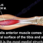
Arises from the upper one-third of the posterior surface of the tibia.
-
Originates from the middle one-third of the medial border of the tibial shaft.
-
Also arises from:
-
The posterior surface of the head of the fibula
-
The upper one-third of the posterior surface of the fibular shaft
-
-
Additional origin from a fibrous arch between the tibia and fibula, known as the soleal arch.
INSERTION
-
The soleus muscle joins the gastrocnemius muscle.
-
Together, they form the Achilles tendon.
-
The Achilles tendon inserts into the posterior surface of the calcaneus.
INNERVATION
-
Supplied by the tibial nerve, with nerve roots S1 and S2.
-
The tibial nerve passes through the fibrous arch of the soleus muscle.
FUNCTION
-
Primary plantar flexor of the ankle joint.
-
Plays a crucial role in:
-
Standing posture
-
Walking and running
-
Preventing forward sway of the body during stance
-
CLINICAL CORRELATIONS
1. Compartment Syndrome
-
A surgical emergency.
-
Characterized by increased pressure within a closed muscle compartment.
-
Leads to compromised microcirculation and tissue ischemia.
-
If untreated, may result in muscle necrosis and permanent disability.
Fasciotomy
-
Performed using a posteromedial incision, placed approximately 2 centimeters posterior to the medial border of the tibia.
-
Allows decompression of:
-
The superficial posterior compartment
-
The deep posterior compartment
-
-
Access to the deep posterior compartment is achieved by releasing the soleus muscle from its tibial attachment.
-
Care must be taken to protect the saphenous nerve.
2. Achilles Tendinitis
-
Caused by overuse and repetitive microtrauma.
-
Results in:
-
Pain
-
Swelling
-
Degenerative changes within the Achilles tendon
-
3. Retrocalcaneal Bursitis
-
Inflammation of the bursa located between the anterior surface of the Achilles tendon and the posterior calcaneus.
-
A common cause of posterior ankle pain, especially in athletes.
4. Achilles Tendon Rupture
-
Rupture commonly occurs proximal to the calcaneal insertion.
-
Blood supply is derived from:
-
Posterior tibial artery
-
Peroneal artery
-
Watershed Zone
-
A region with relatively poor blood supply.
-
Located approximately 2 to 6 centimeters proximal to the calcaneal insertion.
-
This zone is particularly prone to tendon rupture.
Clinical Test
Thompson Test
-
The examiner squeezes the calf muscle.
-
Normal response: plantar flexion of the foot.
-
Complete Achilles tendon rupture:
-
Absence of ankle movement during calf squeeze
-
Indicates loss of tendon continuity
-
DIFFERENTIATING TIGHTNESS
Gastrocnemius Tightness
-
Ankle dorsiflexion is limited when the knee is extended.
-
Gastrocnemius spans the knee joint.
-
Knee flexion relaxes the muscle and improves ankle dorsiflexion.
-
Increased dorsiflexion with knee flexion indicates gastrocnemius tightness.
Achilles Tendon Tightness
-
Ankle dorsiflexion remains equally restricted with:
-
Knee extension
-
Knee flexion
-
-
Indicates contracture or tightness of the Achilles tendon.
-
Degree of dorsiflexion does not change with knee position.
CLINICAL IMPORTANCE
-
The soleus muscle is essential for ankle stability and endurance activities.
-
Pathology involving the soleus or Achilles tendon significantly affects gait and athletic performance.
-
Understanding its anatomy is critical in managing compartment syndrome, tendon injuries, and posterior leg pain.

Leave a Reply