• Skip to main content
  • Skip to secondary menu
  • Skip to primary sidebar
OrthopaedicPrinciples.com

OrthopaedicPrinciples.com

Integrating Principles and Evidence

Integrating Principles and Evidence

  • Home
  • Editorial Board
  • Our Books
    • Evidence Based Orthopaedic Principles
  • Courses
  • Exams
  • Reviews
  • Live Program
  • Contact

Anatomy of the Pelvis-Basics

Courtesy: Prof Nabil Ebraheim, University of Toledo, Ohio, USA

Bony Anatomy of the Pelvis

The pelvis is a ring-like bony structure that connects the spine to the lower limbs and supports pelvic organs.

Components of the Pelvis

  1. Hip Bones (Innominate Bones) – Two large bones, each formed by fusion of:

    • Ilium

    • Ischium

    • Pubis

  2. Sacrum

    • Triangular bone at the base of the spine

    • Formed by fusion of five sacral vertebrae

    • Transfers body weight to the pelvis

  3. Coccyx

    • Terminal part of the vertebral column

    • Composed of fused coccygeal vertebrae

    • Serves as attachment for ligaments and muscles


Ligaments of the Pelvis

Pelvic ligaments provide stability, limit excessive motion, and support pelvic organs by connecting the sacrum, ilium, ischium, and pubis.


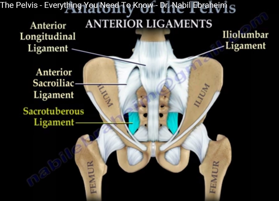
Anterior Pelvic Ligaments

  1. Anterior Longitudinal Ligament

  2. Anterior Sacroiliac Ligament

  3. Sacrotuberous Ligament

  4. Iliofemoral Ligament

  5. Iliolumbar Ligament

  6. Inguinal Ligament

  7. Sacrospinous Ligament

  8. Obturator Membrane


Posterior Pelvic Ligaments

  1. Dorsal Sacroiliac Ligament

    • Major stabilizer of the sacroiliac joint

  2. Supraspinous Ligament

  3. Sacrotuberous Ligament

  4. Sacrospinous Ligament

  5. Iliolumbar Ligament

  6. Articular Capsule of the Hip Joint

  7. Deep Dorsal Sacrococcygeal Ligament

  8. Superficial Dorsal Sacrococcygeal Ligament

  9. Arcuate Pubic Ligament


Muscles Present in the Pelvis

These muscles contribute to hip movement, pelvic stability, and rotation of the lower limb.

  1. Piriformis

  2. Superior Gemellus

  3. Obturator Internus

  4. Inferior Gemellus

  5. Quadratus Femoris

  6. Obturator Externus


Arteries of the Pelvis

Major blood supply to pelvic organs and lower limb originates from iliac vessels.

  1. Common Iliac Artery

  2. External Iliac Artery

  3. Internal Iliac Artery

  4. Pudendal Artery


Veins of the Pelvis

Venous drainage mirrors the arterial system.

  1. External Iliac Vein

  2. Internal Iliac Vein

  3. Common Iliac Vein

  4. Deep Circumflex Iliac Vein

  5. Lateral Sacral Vein


Nerves of the Pelvis

Pelvic nerves supply the lower limb, pelvic floor, and perineum.

  1. Pudendal Nerve

  2. Obturator Nerve

  3. Iliohypogastric Nerve

  4. Ilioinguinal Nerve

  5. Genitofemoral Nerve

  6. Lateral Cutaneous Nerve of Thigh

  7. Superior Gluteal Nerve

  8. Sciatic Nerve

  9. Superior Cluneal Nerves


Joints of the Pelvis

Pelvic joints provide a balance between stability and limited mobility.

  1. Sacroiliac Joint

  2. Sacrococcygeal Joint

  3. Lumbosacral Joint

  4. Pubic Symphysis

  5. Hip Joint


Key Take-Home Points

  • The pelvis acts as a load-bearing ring

  • Stability is mainly ligamentous rather than muscular

  • Internal iliac vessels are the primary supply to pelvic organs

  • Pelvic nerves are clinically important in trauma, childbirth, and surgery

  • Hip joint is the most mobile joint of the pelvis

Pelvis anatomy

Post Views: 4,567

Related Posts

  • Neuronal Anatomy Basics

    Courtesy: Prof Nabile Ebraheim, Professor of Orthopaedics and Traumatology, University of Toledo, Ohio, United States…

  • Pelvic fracture Basics

    Courtesy: Dr Saqib Rehman, MD Associate Professor Director of Orthopaedic Trauma Temple University, Philadelphia Pennsylvania,…

  • Lumbar Plexus Anatomy

    Courtesy: Prof Nabil Ebraheim, University of Toledo, Ohio, USA

Reader Interactions

Leave a Reply

Your email address will not be published. Required fields are marked *

Primary Sidebar

Follow Us

instagram slideshare

Categories

  • -Applied Anatomy
  • -Approaches
  • -Basic Sciences
  • -Cartilage & Meniscus
  • -Classifications
  • -Examination
  • -Foot and Ankle
  • -Foot and Ankle Trauma
  • -FRCS(Tr and Orth) tutorials
  • -Gait
  • -Hand and Wrist
  • -Hand and Wrist Trauma
  • -Hand Infections
  • -Hip and Knee
  • -Hip Preservation
  • -Infections
  • -Joint Reconstruction
  • -Knee Arthroplasty
  • -Knee Preservation
  • -Metabolic Disorders
  • -Oncology
  • -OrthoBiologics
  • -OrthoPlastic
  • -Paediatric Orthopaedics
  • -Paediatric Trauma
  • -Patellofemoral Joint
  • -Pelvis
  • -Peripheral Nerves
  • -Principles
  • -Principles of Surgery
  • -Radiology
  • -Rheumatology
  • -Shoulder and Elbow
  • -Shoulder and Elbow Arthroplasty
  • -Spine Deformity
  • -Spine Oncology
  • -Spine Trauma
  • -Spine, Pelvis & Neurology
  • -Sports Ankle and Foot
  • -Sports Elbow
  • -Sports Knee
  • -Sports Medicine
  • -Sports Medicine Hip
  • -Sports Shoulder
  • -Sports Wrist
  • -Statistics
  • -Technical Tip
  • -Technology in Orth
  • -Trauma
  • -Trauma (Upper Limb)
  • -Trauma Life Support
  • -Trauma Reconstruction
  • Book Shelf
  • Book Shelf Medical
  • Careers
  • Case Studies and Free Papers
  • DNB Ortho
  • Evidence Based Orthopaedic Principles
  • Evidence Based Orthopaedics
  • Exam Corner
  • Fellowships
  • Guest Editor
  • Guest Reviews
  • Image Quiz
  • Instructional Course Lectures
  • Journal Club
  • MCQs
  • Meetings and Courses
  • Multimedia
  • News and Blog
  • Plaster Techniques
  • Podcasts
  • Public Health
  • Rehabilitation
  • Research
  • Shorts and Reels
Copyright@orthopaedicprinciples.com. All right rerserved.