• Skip to main content
  • Skip to secondary menu
  • Skip to primary sidebar
OrthopaedicPrinciples.com

OrthopaedicPrinciples.com

Integrating Principles and Evidence

Integrating Principles and Evidence

  • Home
  • Editorial Board
  • Our Books
    • Evidence Based Orthopaedic Principles
  • Courses
  • Exams
  • Reviews
  • Live Program
  • Contact

Anatomy Of The Gastrocnemius Muscle

Courtesy: Prof Nabil Ebraheim, University of Toledo, Ohio, USA
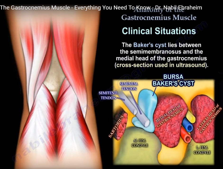
Video describes The Anatomy of Gastrocnemius Muscle. It arises from femur and crosses the knee joint and Ankle joint.It is part of the three Superficial Flexors in the leg. The Gastrocnemius Muscle has 2 heads, one medial and one lateral head. The Medial head originates from the posterior surface of femur above the Medial Femoral condyle while the lateral head originates from the Lateral surface of lateral Femoral condyle. The two bellies of Gastrocnemius Muscle end in a tendon which joins with the tendon of soleus muscle to form together the tendoachilles or tendocalcaneus which inserts into the middle third of the posterior surface of calcaneus. This Muscle is innervated by tibial nerve (S1,S2) and it is supplied by Sural Branch of Popliteal artery. Together with the soleus muscle, this Muscle is a powerful plantar flexor of Ankle. Along with that it also helps in knee flexion.

Post Views: 8,059

Related Posts

  • Popliteus Muscle

    Courtesy : Prof Nabile Ebraheim, University of Toledo, Ohio, USA Popliteus Muscle A triangular muscle…

  • Muscle Anatomy of the Trunk

    Courtesy: Prof Nabil Ebraheim, University of Toledo, Ohio, USA

  • Tibialis Anterior Muscle Anatomy

    ?   ANATOMY OF TIBIALIS ANTERIOR MUSCLE The leg has four muscle compartments Anterior Lateral…

Reader Interactions

Leave a Reply

Your email address will not be published. Required fields are marked *

Primary Sidebar

Follow Us

instagram slideshare

Categories

  • -Applied Anatomy
  • -Approaches
  • -Basic Sciences
  • -Cartilage & Meniscus
  • -Classifications
  • -Examination
  • -Foot and Ankle
  • -Foot and Ankle Trauma
  • -FRCS(Tr and Orth) tutorials
  • -Gait
  • -Hand and Wrist
  • -Hand and Wrist Trauma
  • -Hand Infections
  • -Hip and Knee
  • -Hip Preservation
  • -Infections
  • -Joint Reconstruction
  • -Knee Arthroplasty
  • -Knee Preservation
  • -Metabolic Disorders
  • -Oncology
  • -OrthoBiologics
  • -OrthoPlastic
  • -Paediatric Orthopaedics
  • -Paediatric Trauma
  • -Patellofemoral Joint
  • -Pelvis
  • -Peripheral Nerves
  • -Principles
  • -Principles of Surgery
  • -Radiology
  • -Rheumatology
  • -Shoulder and Elbow
  • -Shoulder and Elbow Arthroplasty
  • -Spine Deformity
  • -Spine Oncology
  • -Spine Trauma
  • -Spine, Pelvis & Neurology
  • -Sports Ankle and Foot
  • -Sports Elbow
  • -Sports Knee
  • -Sports Medicine
  • -Sports Medicine Hip
  • -Sports Shoulder
  • -Sports Wrist
  • -Statistics
  • -Technical Tip
  • -Technology in Orth
  • -Trauma
  • -Trauma (Upper Limb)
  • -Trauma Life Support
  • -Trauma Reconstruction
  • Book Shelf
  • Book Shelf Medical
  • Careers
  • Case Studies and Free Papers
  • DNB Ortho
  • Evidence Based Orthopaedic Principles
  • Evidence Based Orthopaedics
  • Exam Corner
  • Fellowships
  • Guest Editor
  • Guest Reviews
  • Image Quiz
  • Instructional Course Lectures
  • Journal Club
  • MCQs
  • Meetings and Courses
  • Multimedia
  • News and Blog
  • Plaster Techniques
  • Podcasts
  • Public Health
  • Rehabilitation
  • Research
  • Shorts and Reels
Copyright@orthopaedicprinciples.com. All right rerserved.