• Skip to main content
  • Skip to secondary menu
  • Skip to primary sidebar
OrthopaedicPrinciples.com

OrthopaedicPrinciples.com

Integrating Principles and Evidence

Integrating Principles and Evidence

  • Home
  • Editorial Board
  • Our Books
    • Evidence Based Orthopaedic Principles
  • Courses
  • Exams
  • Reviews
  • Live Program
  • Contact

Anatomy of the Flexor Hallucis Longus

Courtesy : Prof Nabile Ebraheim, University of Toledo, Ohio, USA

FLEXOR HALLUCIS LONGUS
• One Of The Deep Muscles Of Posterior Compartment Of The Leg
• FHL Muscle Spans Down The Calf To The Side Of Ankle And Into Foot
• Muscle Fibers – Lowest Origin In Leg And Some Fibers Almost At The Ankle

ORIGIN AND INSERTION
• FHL Muscle Arises From Posterior Surface On Inferior 2/3rds Of Fibula
• As Muscle Extends Down Into Plantar Aspect Of The Foot , Tendon Inserts Into Distal Phalanx Of Large Toe.
• FHL Muscle Is Largest And Strongest Deep Muscle Of Leg’s Posterior Section.
• There Are Two Sesamoid Bones At Level Of The MCP Joint , One Tibial And One Fibular- That Act Like Pulleys For The Flexor Tendons and are embedded in The Flexor Hallucis Brevis Muscle
• Sesamoids Are Important To The Big Toe Region By Absorbing Weight Bearing Pressure And Reducing Friction On Metatarsal Head.
• Also They Protect The Flexor Hallucis Longus Tendon As It Glides Between Two Sesamoid Bones

FUNCTION & INNERVATION
• FHL Flexes Joints Of Big Toe, And Also Role In Plantar Flexion Of Ankle Joint
• Tibial Nerve Provides Innervation To The Flexor Hallucis Longus Muscle
• Blood Supply From Peroneal Artery(Muscular Branch) And Posterior Tibial Artery (Peroneal Branch)

ARRANGEMENT OF TENDONS
• The FHL tendon has long course from its muscle origin to its insertion
• The tendon is vulnerable to wear and tear , friction and inflammation at certain anatomic locations
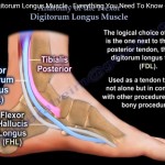
• At the ankle FHL located posterior and lateral to FDL and in between posterior talar processes
• The tendon lies most lateral in medial ankle compartment
• The tendon passes in its own tunnel beneath the sustentaculum tali

• Tendinopathy is common at the tibiotalar joint as the tendon curves over the posterior talus
• Flexor hallucis tendonitis mostly happens in ballet dancers and those require excessive plantar flexion at ankle
• Distal disease of hallux sesamoids is affected next, followed by area of knot of henry
• As tendons of FHL and FDL curve under medial malleolus and talus , they begin to converge and eventually cross
• At this point KNOT OF HENRY fibrous slip connects the FDL with FHL and tendon sheath communicates allowing spreading of inflammation
• Because of the intersection of two tendons , transection of digitorum proximal to the KNOT OF HENRY to correct the tibialis posterior dysfunction could result in retention of function of hallux and lesser digits

Post Views: 2,753

Related Posts

  • Anatomy of Flexor Digitorum Longus

    ? Courtesy: Prof Nabil Ebraheim, University of Toledo, Ohio, USA Anatomy of Flexor Digitorum Longus…

  • Anatomy of the Flexor Pollicis Longus

    Courtesy: Prof Nabil Ebraheim, University of Toledo, Ohio, USA Video describes the Anatomy of Flexor…

  • Flexor Tendon Anatomy

    Courtesy: Prof Nabil Ebraheim The ability to flex the fingers consists of a system of…

Reader Interactions

Leave a Reply

Your email address will not be published. Required fields are marked *

Primary Sidebar

Follow Us

instagram slideshare

Categories

  • -Applied Anatomy
  • -Approaches
  • -Basic Sciences
  • -Cartilage & Meniscus
  • -Classifications
  • -Examination
  • -Foot and Ankle
  • -Foot and Ankle Trauma
  • -FRCS(Tr and Orth) tutorials
  • -Gait
  • -Hand and Wrist
  • -Hand and Wrist Trauma
  • -Hand Infections
  • -Hip and Knee
  • -Hip Preservation
  • -Infections
  • -Joint Reconstruction
  • -Knee Arthroplasty
  • -Knee Preservation
  • -Metabolic Disorders
  • -Oncology
  • -OrthoBiologics
  • -OrthoPlastic
  • -Paediatric Orthopaedics
  • -Paediatric Trauma
  • -Patellofemoral Joint
  • -Pelvis
  • -Peripheral Nerves
  • -Principles
  • -Principles of Surgery
  • -Radiology
  • -Rheumatology
  • -Shoulder and Elbow
  • -Shoulder and Elbow Arthroplasty
  • -Spine Deformity
  • -Spine Oncology
  • -Spine Trauma
  • -Spine, Pelvis & Neurology
  • -Sports Ankle and Foot
  • -Sports Elbow
  • -Sports Knee
  • -Sports Medicine
  • -Sports Medicine Hip
  • -Sports Shoulder
  • -Sports Wrist
  • -Statistics
  • -Technical Tip
  • -Technology in Orth
  • -Trauma
  • -Trauma (Upper Limb)
  • -Trauma Life Support
  • -Trauma Reconstruction
  • Book Shelf
  • Book Shelf Medical
  • Careers
  • Case Studies and Free Papers
  • DNB Ortho
  • Evidence Based Orthopaedic Principles
  • Evidence Based Orthopaedics
  • Exam Corner
  • Fellowships
  • Guest Editor
  • Guest Reviews
  • Image Quiz
  • Instructional Course Lectures
  • Journal Club
  • MCQs
  • Meetings and Courses
  • Multimedia
  • News and Blog
  • Plaster Techniques
  • Podcasts
  • Public Health
  • Rehabilitation
  • Research
  • Shorts and Reels
Copyright@orthopaedicprinciples.com. All right rerserved.