• Skip to main content
  • Skip to secondary menu
  • Skip to primary sidebar
OrthopaedicPrinciples.com

OrthopaedicPrinciples.com

Integrating Principles and Evidence

Integrating Principles and Evidence

  • Home
  • Editorial Board
  • Our Books
    • Evidence Based Orthopaedic Principles
  • Courses
  • Exams
  • Reviews
  • Live Program
  • Contact

Anatomy of Quadratus Femoris

Courtesy: Prof Nabil Ebraheim, University of Toledo, Ohio, USA

ANATOMY OF QUADRATUS FEMORIS MUSCLE

ORIGIN

  • From the lateral margin of ischial tuberosity

INSERTION

  • Into the quadrate tubercle on the intertrochanteric crest between the greater and lesser trochanter

ACTION

  • External rotation of the hip and also helps in adduction of thigh

NERVE SUPPLY

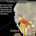
  • The nerve from sacral plexus called the nerve to quadratus femoris(L5, S1)

RELATIONS

  • Medial Femoral Circumflex Artery courses anterior to the superior edge of quadratus muscle
  • This artery can be identified in the space between the superior edge of quadratus femoris and inferior gemellus

CLINICAL SIGNIFICANCE

  • The detachment of quadratus femoris from femur during the posterior approach to hip results in profuse bleeding from the branch of the medial femoral circumflex artery.

Post Views: 10,372

Related Posts

  • Anatomy of Pronator Quadratus

    Courtesy: Prof Nabile Ebraheim, University of Toledo, Ohio, USA Anatomy of Pronator Quadratus  it is…

  • Anatomy of Rectus Femoris Muscle

    Courtesy: Prof Nabil Ebrhaeim, University of Toledo, Ohio, USA Video describes Anatomy of Rectus Femoris…

  • Sciatica Anatomy

    Courtesy: Prof Nabil Ebraheim University of Toledo, Ohio, USA Anatomy of sciatica The sciatic nerve…

Reader Interactions

Leave a Reply

Your email address will not be published. Required fields are marked *

Primary Sidebar

Follow Us

instagram slideshare

Categories

  • -Applied Anatomy
  • -Approaches
  • -Basic Sciences
  • -Cartilage & Meniscus
  • -Classifications
  • -Examination
  • -Foot and Ankle
  • -Foot and Ankle Trauma
  • -FRCS(Tr and Orth) tutorials
  • -Gait
  • -Hand and Wrist
  • -Hand and Wrist Trauma
  • -Hand Infections
  • -Hip and Knee
  • -Hip Preservation
  • -Infections
  • -Joint Reconstruction
  • -Knee Arthroplasty
  • -Knee Preservation
  • -Metabolic Disorders
  • -Oncology
  • -OrthoBiologics
  • -OrthoPlastic
  • -Paediatric Orthopaedics
  • -Paediatric Trauma
  • -Patellofemoral Joint
  • -Pelvis
  • -Peripheral Nerves
  • -Principles
  • -Principles of Surgery
  • -Radiology
  • -Rheumatology
  • -Shoulder and Elbow
  • -Shoulder and Elbow Arthroplasty
  • -Spine Deformity
  • -Spine Oncology
  • -Spine Trauma
  • -Spine, Pelvis & Neurology
  • -Sports Ankle and Foot
  • -Sports Elbow
  • -Sports Knee
  • -Sports Medicine
  • -Sports Medicine Hip
  • -Sports Shoulder
  • -Sports Wrist
  • -Statistics
  • -Technical Tip
  • -Technology in Orth
  • -Trauma
  • -Trauma (Upper Limb)
  • -Trauma Life Support
  • -Trauma Reconstruction
  • Book Shelf
  • Book Shelf Medical
  • Careers
  • Case Studies and Free Papers
  • DNB Ortho
  • Evidence Based Orthopaedic Principles
  • Evidence Based Orthopaedics
  • Exam Corner
  • Fellowships
  • Guest Editor
  • Guest Reviews
  • Image Quiz
  • Instructional Course Lectures
  • Journal Club
  • MCQs
  • Meetings and Courses
  • Multimedia
  • News and Blog
  • Plaster Techniques
  • Podcasts
  • Public Health
  • Rehabilitation
  • Research
  • Shorts and Reels
Copyright@orthopaedicprinciples.com. All right rerserved.