• Skip to main content
  • Skip to secondary menu
  • Skip to primary sidebar
OrthopaedicPrinciples.com

OrthopaedicPrinciples.com

Integrating Principles and Evidence

Integrating Principles and Evidence

  • Home
  • Editorial Board
  • Our Books
    • Evidence Based Orthopaedic Principles
  • Courses
  • Exams
  • Reviews
  • Live Program
  • Contact

Anatomy of Gracilis Muscle

Courtesy: Prof Nabil Ebraheim, University of Toledo, Ohio, USA


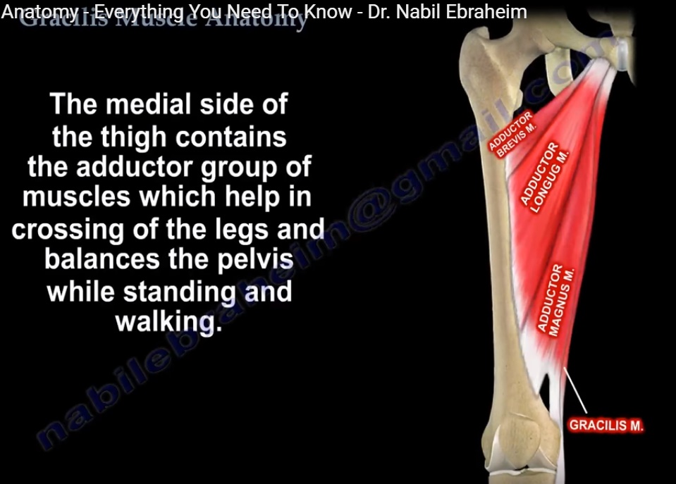
GRACILIS MUSCLE ANATOMY

  • It is the most superficial muscle on the medial side of thigh
  • The tendon of gracilis muscle is easily palpable in the inguinal region together with adductor longus muscle.
  • Medial side of the thigh contains the adductor group of muscles which help in crossing of the legs and balances the pelvis while standing and walking.
  • It is the only muscle that crosses 2 joints. It provides a reliable flap for coverage. Most common donor muscle for free muscle transfer procedure.
  • Origin : outer surface of ischiopubic ramus.
  • Insertion: Upper medial part of tibial shaft below the sartorius muscle. The area is called Pes Ansérines.
  • It is the only muscle of the medial aspect of the adductors of thigh that doesn’t insert into the line aspera of femur.
  • Innervation: Anterior branch of the obturator nerve
  • Arises from the lumbar plexus L2, L3, L4.
  • It passes through the obturator foramen to reach the adductor muscles.
  • Function : adducts the hip, flexes and internally rotates the knee.

Gracilis anatomy

Post Views: 7,365

Related Posts

  • Popliteus Muscle

    Courtesy : Prof Nabile Ebraheim, University of Toledo, Ohio, USA Popliteus Muscle A triangular muscle…

  • Tibialis Anterior Muscle Anatomy

    ?   ANATOMY OF TIBIALIS ANTERIOR MUSCLE The leg has four muscle compartments Anterior Lateral…

  • Muscle Anatomy of the Trunk

    Courtesy: Prof Nabil Ebraheim, University of Toledo, Ohio, USA

Reader Interactions

Comments

  1. uttam pal says

    at

    How to relax gracilis muscle contracture in hemiplegic patient?

Leave a Reply

Your email address will not be published. Required fields are marked *

Primary Sidebar

Follow Us

instagram slideshare

Categories

  • -Applied Anatomy
  • -Approaches
  • -Basic Sciences
  • -Cartilage & Meniscus
  • -Classifications
  • -Examination
  • -Foot and Ankle
  • -Foot and Ankle Trauma
  • -FRCS(Tr and Orth) tutorials
  • -Gait
  • -Hand and Wrist
  • -Hand and Wrist Trauma
  • -Hand Infections
  • -Hip and Knee
  • -Hip Preservation
  • -Infections
  • -Joint Reconstruction
  • -Knee Arthroplasty
  • -Knee Preservation
  • -Metabolic Disorders
  • -Oncology
  • -OrthoBiologics
  • -OrthoPlastic
  • -Paediatric Orthopaedics
  • -Paediatric Trauma
  • -Patellofemoral Joint
  • -Pelvis
  • -Peripheral Nerves
  • -Principles
  • -Principles of Surgery
  • -Radiology
  • -Rheumatology
  • -Shoulder and Elbow
  • -Shoulder and Elbow Arthroplasty
  • -Spine Deformity
  • -Spine Oncology
  • -Spine Trauma
  • -Spine, Pelvis & Neurology
  • -Sports Ankle and Foot
  • -Sports Elbow
  • -Sports Knee
  • -Sports Medicine
  • -Sports Medicine Hip
  • -Sports Shoulder
  • -Sports Wrist
  • -Statistics
  • -Technical Tip
  • -Technology in Orth
  • -Trauma
  • -Trauma (Upper Limb)
  • -Trauma Life Support
  • -Trauma Reconstruction
  • Book Shelf
  • Book Shelf Medical
  • Careers
  • Case Studies and Free Papers
  • DNB Ortho
  • Evidence Based Orthopaedic Principles
  • Evidence Based Orthopaedics
  • Exam Corner
  • Fellowships
  • Guest Editor
  • Guest Reviews
  • Image Quiz
  • Instructional Course Lectures
  • Journal Club
  • MCQs
  • Meetings and Courses
  • Multimedia
  • News and Blog
  • Plaster Techniques
  • Podcasts
  • Public Health
  • Rehabilitation
  • Research
  • Shorts and Reels
Copyright@orthopaedicprinciples.com. All right rerserved.