Courtesy: Prof Nabil Ebraheim, University of Toledo, Ohio, USA
Overview
- The brachial plexus is the nerve network that supplies motor and sensory innervation to the upper limb.
- It is formed by the anterior rami of five spinal nerves.
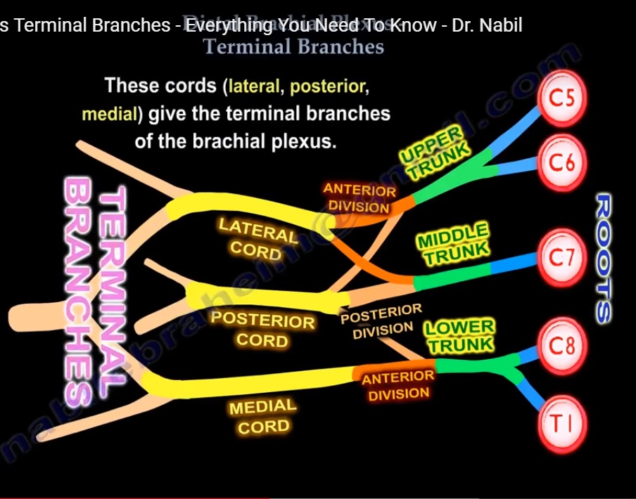
- These spinal nerve roots combine and reorganize to form trunks, divisions, cords, and finally the terminal nerves of the upper limb.
Formation of the Brachial Plexus
Nerve Roots
- The brachial plexus originates from five spinal nerve roots.
- These roots are:
- C5
- C6
- C7
- C8
- T1
- These roots represent the ventral rami of the spinal nerves that supply the upper limb.
Trunks
The nerve roots combine to form three trunks:
- Upper trunk
- Formed by the union of C5 and C6 roots.
- Middle trunk
- Continuation of the C7 root alone.
- Lower trunk
- Formed by the union of C8 and T1 roots.
Divisions
- Each trunk divides into two branches:
- Anterior division
- Posterior division
- Therefore, the brachial plexus contains:
- Three anterior divisions
- Three posterior divisions
Cords
The divisions reorganize to form three cords.
These cords are named according to their relationship to the axillary artery deep to the pectoralis minor muscle.
Posterior Cord
- Formed by the union of all three posterior divisions.
- Lies posterior to the axillary artery.
Lateral Cord
- Formed by the anterior divisions of the upper and middle trunks.
- Lies lateral to the axillary artery.
Medial Cord
- Formed by the anterior division of the lower trunk.
- Lies medial to the axillary artery.
Terminal Branches of the Brachial Plexus
The cords give rise to the major terminal nerves that supply the upper limb.
Axillary Nerve
- Arises from the posterior cord.
- Supplies the deltoid and teres minor muscles.
- Provides sensory supply to the skin over the lateral shoulder region.
Radial Nerve
- Continuation of the posterior cord after giving the axillary nerve.
- Supplies the extensor muscles of the arm and forearm.
- Provides sensory supply to the posterior aspect of the upper limb.
Musculocutaneous Nerve
- Continuation of the lateral cord.
- Supplies the flexor muscles of the anterior compartment of the arm.
- Continues distally as a sensory nerve of the lateral forearm.
Median Nerve
- Formed by the union of two roots:
- Lateral root from the lateral cord
- Medial root from the medial cord
- These roots unite on the anterior surface of the axillary artery.
Functions include:
- Motor supply to most flexor muscles of the forearm.
- Motor supply to some intrinsic muscles of the hand.
- Sensory supply to the lateral aspect of the palm and fingers.
Ulnar Nerve
- Continuation of the medial cord after contributing to the median nerve.
- Supplies most intrinsic muscles of the hand.
- Provides sensory supply to the medial side of the hand and fingers.
Important Anatomical Arrangement
- The terminal nerves in the axilla form a characteristic “M” shaped pattern over the axillary artery.
The three limbs of this pattern are:
- Lateral limb: Musculocutaneous nerve
- Central limb: Median nerve
- Medial limb: Ulnar nerve
This arrangement is an important anatomical landmark during surgical and clinical examination of the axilla.

Leave a Reply