• Skip to main content
  • Skip to secondary menu
  • Skip to primary sidebar
OrthopaedicPrinciples.com

OrthopaedicPrinciples.com

Integrating Principles and Evidence

Integrating Principles and Evidence

  • Home
  • Editorial Board
  • Our Books
    • Evidence Based Orthopaedic Principles
  • Courses
  • Exams
  • Reviews
  • Live Program
  • Contact

Anatomy and Contents of the Popliteal fossa

Courtesy: Prof Nabil Ebraheim, University of Toledo, Ohio, USA

Popliteal fossa is a shallow depression located at the back of the knee joint , bounded by biceps femoris superolaterally ,semimembranosus and semitendinosus superomedially . Inferior boundary of this space is made by the medial and lateral heads of gastrocnemius muscle .
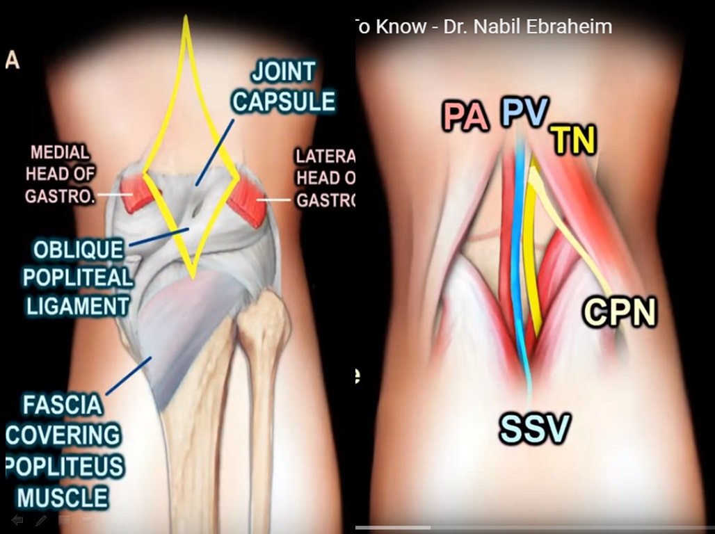
Coming to the base of the popliteal fossa :popliteal surface of the femur ,capsule of the knee joint , oblique popliteal ligament,and the strong fascia covering the popliteus muscle constitute the floor.Popliteal artery, popliteal vein ,small saphenous vein ,tibial nerve and the common peroneal nerve are the structures located within the popliteal fossa in medial to lateral direction. Skin and the deep popliteal fascia form the roof .
Baker’s cyst otherwise called as popliteal cyst is an important clinical entity related to the popliteal fossa ,located between semimembranosus and the medial gastrocnemius ,it is commonly caused by knee arthritis or a meniscal tear.The cyst is connected to the knee joint through a valvular opening .knee effusion from intra articular pathology allows the fluid to go through the valve to the cyst in one direction.

Post Views: 10,306

Related Posts

  • Popliteal Fossa-Anatomy

    Courtesy: Prof Nabil Ebraheim, University of Toledo, Ohio, USA

  • Anatomy of the Popliteal Fossa

    Courtesy: Harry Benjamin Laing, MRCS, Ortho M8, FRCS(Tr and Orth) Tutorials

  • Anatomy of Cubital Fossa

    Courtesy: Prof Nabil Ebraheim, University of Toledo, Ohio, USA

Reader Interactions

Leave a Reply

Your email address will not be published. Required fields are marked *

Primary Sidebar

Follow Us

instagram slideshare

Categories

  • -Applied Anatomy
  • -Approaches
  • -Basic Sciences
  • -Cartilage & Meniscus
  • -Classifications
  • -Examination
  • -Foot and Ankle
  • -Foot and Ankle Trauma
  • -FRCS(Tr and Orth) tutorials
  • -Gait
  • -Hand and Wrist
  • -Hand and Wrist Trauma
  • -Hand Infections
  • -Hip and Knee
  • -Hip Preservation
  • -Infections
  • -Joint Reconstruction
  • -Knee Arthroplasty
  • -Knee Preservation
  • -Metabolic Disorders
  • -Oncology
  • -OrthoBiologics
  • -OrthoPlastic
  • -Paediatric Orthopaedics
  • -Paediatric Trauma
  • -Patellofemoral Joint
  • -Pelvis
  • -Peripheral Nerves
  • -Principles
  • -Principles of Surgery
  • -Radiology
  • -Rheumatology
  • -Shoulder and Elbow
  • -Shoulder and Elbow Arthroplasty
  • -Spine Deformity
  • -Spine Oncology
  • -Spine Trauma
  • -Spine, Pelvis & Neurology
  • -Sports Ankle and Foot
  • -Sports Elbow
  • -Sports Knee
  • -Sports Medicine
  • -Sports Medicine Hip
  • -Sports Shoulder
  • -Sports Wrist
  • -Statistics
  • -Technical Tip
  • -Technology in Orth
  • -Trauma
  • -Trauma (Upper Limb)
  • -Trauma Life Support
  • -Trauma Reconstruction
  • Book Shelf
  • Book Shelf Medical
  • Careers
  • Case Studies and Free Papers
  • DNB Ortho
  • Evidence Based Orthopaedic Principles
  • Evidence Based Orthopaedics
  • Exam Corner
  • Fellowships
  • Guest Editor
  • Guest Reviews
  • Image Quiz
  • Instructional Course Lectures
  • Journal Club
  • MCQs
  • Meetings and Courses
  • Multimedia
  • News and Blog
  • Plaster Techniques
  • Podcasts
  • Public Health
  • Rehabilitation
  • Research
  • Shorts and Reels
Copyright@orthopaedicprinciples.com. All right rerserved.