
Courtesy: Mary Mulcahey MD, Professor, Loyola University School of Medicine, Chicago, USA
Topic / Overview
Acromioclavicular (AC) joint injuries represent a spectrum of shoulder trauma ranging from mild ligamentous sprains to complete dislocations with multidirectional instability. Management encompasses conservative modalities, primary repair, and complex anatomic or non-anatomic reconstructions.
Background / Problems
-
Epidemiology:
-
Accounts for approximately 12% of all shoulder injuries.
-
5 to 10 times more common in males than females.
-
Predominantly occurs in the first three decades of life.
-
High-risk activities: Contact and collision sports (football, rugby, hockey) and fall-risk activities (bicycling, skiing).
-
-
Mechanism of Injury:
-
Direct trauma (most common): Direct blow to the lateral aspect of the shoulder with the arm in an adducted position.
-
Severe direct forces: Can lead to concomitant avulsion/tearing of the deltoid and trapezius muscle attachments from the clavicle.
-
Uncommon indirect mechanisms: Traction forces (substantial pull through the upper extremity) or laterally directed forces coupled with scapular external rotation.
-
-
Functional Anatomy & Biomechanics:
-
Joint Type: Diarthrodial gliding joint between the medial facet of the acromion and the lateral distal clavicle.
-
Motion Dynamics: Assists shoulder abduction and flexion. During arm elevation, the clavicle rotates 40° to 50°, while only 5° to 8° of angular motion occurs at the AC joint itself. Upward clavicular rotation is matched with inferior scapular rotation (synchronous scapuloclavicular motion).
-
AC Joint Capsule & Ligaments (Horizontal Stabilizers):
-
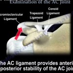
Superior, inferior, and posterior capsule thickenings restrain horizontal translation and posterior rotational forces during scapular protraction.
-
Composed of two distinct bundles: superoposterior (higher structural composition/quality) and anteroinferior (crucial for resisting horizontal displacement).
-
Biomechanical threshold: Sectioning just 50% of the AC capsule reduces resistance to torque across all modalities and significantly increases multidirectional joint motion; complete capsule transection reduces translational resistance force to <25% and rotational resistance force to <10%.
-
-
Coracoclavicular (CC) Ligaments (Vertical Stabilizers & Rotational Restraints):
-
Restrain vertical displacement and scapular internal rotation.
-
Conoid Ligament: Originates from the posterior aspect of the coracoid process and inserts onto the conoid tubercle of the clavicle, 42 to 47 mm medial to the lateral clavicular edge. Confers 60% of the primary restraint to anterosuperior clavicular displacement and rotation.
-
Trapezoid Ligament: Inserts onto the trapezoid line, 20 to 25 mm medial to the distal clavicular edge. Primarily provides resistance against AC joint axial compression.
-
-
-
Associated Pathology:
-
Present in 15% to 18% of AC separations; most commonly SLAP tears and rotator cuff tears.
-
Classification or Concept
1. Rockwood Classification (1984, expanding Tossy [1960s])
-
Type I: Sprain of the AC capsule/ligaments; CC ligaments completely intact. Radiographs demonstrate normal CC distance and no joint widening.
-
Type II: Complete tear of AC ligaments with a sprain or partial tear of the CC ligaments. Radiographs reveal mild vertical subluxation of the distal clavicle.
-
Type III: Complete disruption of both AC and CC ligaments. Radiographs show 25% to 100% vertical displacement compared to the contralateral shoulder.
-
Type IV: Complete disruption of AC and CC ligaments with posterior subluxation/displacement of the clavicle into or through the trapezius muscle (Exam Mnemonic: “Out the back door”).
-
Type V: Complete ligamentous disruption accompanied by extensive stripping of the deltotrapezial fascia; marked vertical displacement (>100% to 300% or severe high displacement; Exam Mnemonic: “High-five”).
-
Type VI: Inferior dislocation of the distal clavicle into the subcoracoid (or subacromial) position (Exam Mnemonic: “Deep six”).
2. ISAKOS Subclassification for Rockwood Type III Injuries
-
Type IIIA (Stable): Horizontally stable distal clavicle. Demonstrates no dynamic horizontal translation; typically responds well to conservative therapy.
-
Type IIIB (Unstable): Horizontally unstable. Evident on cross-body adduction views by distal clavicular overriding; frequently associated with persistent, therapy-resistant scapular dyskinesis that warrants early operative consideration.
3. Scapular Dyskinesis & SICK Scapula Syndrome
-
Alteration of normal scapulothoracic kinematics due to the loss of the AC joint strut/fulcrum function.
-
Dyskinesis Subtypes:
-
Type 1: Prominence of the inferomedial scapular border.
-
Type 2: Prominence of the entire medial scapular border.
-
Type 3: Prominence of the superomedial scapular border.
-
-
SICK Scapula Syndrome: Scapular malposition, Inferior medial border prominence, Coracoid pain/malposition, and dysKinesis of scapular movement.
-
Prevalence in Chronic Type III: Evident in ~70% of patients at rest; two-thirds exhibit inferior border prominence, and 58% develop full SICK scapula syndrome, leading to inferior Constant functional scores.
Indications / Patient Selection
-
Types I and II: Conservative management. Sling immobilization, analgesics/NSAIDs, activity modification, followed by progressive physical therapy. Unrestricted return to contact sports/heavy lifting allowed once full, painless, symmetric motion is regained.
-
Type III (Controversial):
-
First-Line: Trial of non-operative management and rehabilitation.
-
Operative Indications: High-demand overhead throwing athletes, manual laborers, prominent skin tenting threatening soft-tissue integrity, documented horizontal instability (Type IIIB), persistent symptomatic pain, or progressive scapular dyskinesis refractory to physical therapy.
-
-
Types IV, V, and VI: Operative stabilization and ligament reconstruction.
Technique / Principles
Over 150 to 160 surgical techniques have been described, categorized broadly into primary repair, rigid fixation, and ligament reconstruction.
Anatomic CC and AC Ligament Reconstruction (Speaker’s Preferred Technique)
-
Exposure & Coracoid Passage:
-
Open approach; exposure of the distal clavicle and base of the coracoid.
-
A specialized curved/hooked suture-passing device is advanced around the undersurface of the coracoid base to loop passing sutures.
-
-
Bone Tunnel Placement:
-
Clavicular drill holes are placed using a small 2 mm drill bit to minimize bone loss and fracture risk.
-
Tunnels replicate native anatomy: trapezoid tunnel at ~20 mm and conoid tunnel at ~40 mm medial to the distal clavicular margin.
-
-
Implant Passage & Construct Augmentation:
-
Both a soft-tissue allograft (e.g., tendon allograft) and high-strength suture tape are passed beneath the coracoid.
-
Suture tape limbs are routed superiorly through the 2 mm clavicular drill holes.
-
The allograft limbs are directed around the clavicle (one limb passed posteriorly).
-
-
Reduction & Fixation:
-
Manual reduction of the AC joint (applying downward pressure to the clavicle while providing superior axial support at the elbow).
-
Graft limbs are secured over the superior clavicle with heavy non-absorbable/braided suture (e.g., #1 Vicryl or similar).
-
Suture tape limbs are tied tightly over the top of the graft construct, augmenting primary vertical stability.
-
-
Horizontal AC Stabilization:
-
The remaining long limb of the allograft (supplemented with a limb of suture tape) is routed directly across the AC joint onto the acromion and secured into local tissue/capsule to restore anteroposterior stability.
-
-
Soft Tissue Closure:
-
Meticulous, secure repair of the deltotrapezial fascia over the construct, followed by routine layered closure.
-
Distal Clavicle Resection (Optional): Not routinely indicated; reserved selectively when anatomic reduction of the joint is blocked.
-
Surgical Alternatives Described
-
Clavicular Hook Plate: Rigid metallic implant maintaining reduction during healing.
-
Suspensory Fixation: Suture-button/dog-bone cortical button suspensory devices.
-
Kirschner Wires (K-wires): Transfixion pins traversing the acromion into the distal clavicle (largely historical/discouraged).
-
Arthroscopic-Assisted Reconstruction: Emerging technique allowing direct intra-articular inspection and minimally invasive coracoid drilling.
Results / Survivorship / Evidence Cited by the Speaker
-
Radiographic vs. Clinical Course (Types I & II): AJSM study demonstrated that while post-traumatic radiographic changes (distal clavicular osteolysis, CC ligament ossification, local degeneration) are frequent, long-term functional scores remain excellent, with clinically non-significant functional differences compared to the uninjured shoulder.
-
Operative vs. Non-operative Outcomes in Type III:
-
Meta-analyses show no significant differences in strength, pain relief, throwing ability, or secondary osteoarthritis between surgical and non-surgical groups.
-
Surgery provides a superior cosmetic profile (less residual prominence) at the cost of surgical scarring and increased sick leave.
-
Re-dislocation / recurrence rate following operative intervention for Type III: ~14%.
-
-
Comprehensive Systematic Review (20 Studies / 821 References):
-
Favorable outcomes: 80% operative vs. 85% non-operative.
-
Return to work and athletics occurred earlier with non-operative management.
-
Early intervention achieved favorable outcomes in 90%, compared to 70% for delayed surgery.
-
Anatomic reconstruction techniques consistently demonstrated superior functional outcomes compared to non-anatomic procedures.
-
-
Biomechanical Stability:
-
Reconstructing both the CC ligaments and the AC joint capsule yields the highest resistance torque.
-
Adding AC capsule repair/augmentation restores native translational (AP) stability, though residual rotational instability persists under biomechanical testing.
-
-
Return to Sport (RTS):
-
Systematic review data shows an overall RTS rate of 94% to 100% (with 60% to 100% returning to their pre-injury competitive level).
-
Comparable RTS rates observed across Type III and Type V injuries, and across varying operative techniques.
-
Complications / Limitations
-
Hook Plate Morbidity:
-
Associated with the highest overall complication rates alongside K-wires.
-
Key complications: Subacromial osteolysis, acromial erosion/cut-through (particularly in thinner or osteopenic acromia), hardware failure/loss of fixation, and deep wound infection.
-
Mandatory secondary surgery required for plate removal.
-
-
K-Wire Morbidity:
-
High risk of migration, pin site infection, loss of reduction, and catastrophic hardware dislodgement.
-
-
General Reoperation Rates:
-
Unplanned reoperation rate: ~5% for modified Weaver-Dunn procedures; 2% to 3% for hook plates and K-wires (excluding planned plate removal).
-
-
Persistent Biomechanical Weakness:
-
Even combined CC and AC capsule reconstruction constructs remain biomechanically weaker than native, uninjured tissue.
-
-
Subjective Deformity:
-
Surgical patients frequently retain a subtle residual step-off alongside a surgical scar.
-
Comparison with Alternatives
| Feature / Metric | Operative Management | Non-Operative Management |
| Primary Indication | Types IV, V, VI; refractory/unstable Type IIIB; high-demand athletes | Types I, II; uncomplicated Type IIIA |
| Favorable Outcome Rate | ~80% (90% if treated early; 70% if delayed) | ~85% |
| Cosmetic Profile | Superior reduction of prominence; leaves a scar | Persistent bump/step-off; no scar |
| Time to Return (Work/Sport) | Prolonged (rehab-dependent, up to 6 months) | Significantly quicker |
| Shoulder Strength & Pain | Equivalent to non-operative at long-term follow-up | Equivalent to operative at long-term follow-up |
| Scapular Dyskinesis Risk | Lower if anatomically reduced | High in chronic displacement (~70%) |
| Associated Complications | Infection, implant cut-through, osteolysis, failure (~14%) | Residual instability, SICK scapula, cosmetic bump |
Comparison of Reconstructive Implants & Techniques
| Technique / Implant | Advantages | Disadvantages / Complications |
| Anatomic Graft + Suture Tape | Restores vertical and horizontal stability; no routine hardware removal | Technically demanding; requires biological incorporation |
| Clavicular Hook Plate | Provides rigid initial vertical fixation; allows immediate stability | High rate of osteolysis, acromial cut-through/erosion; requires routine hardware removal |
| Kirschner Wires (K-wires) | Historically simple, low upfront cost | Significant complication profile; pin migration; risk of hardware failure |
| Suspensory Cortical Buttons | Minimally invasive/flexible; eliminates mandatory secondary removal | Risk of drill-hole osteolysis or coracoid/clavicle fracture |
| Allograft vs. Autograft | Equivalent clinical outcomes; allograft avoids donor site morbidity | Allografts carry high financial cost and regional availability limitations |
Practical Clinical Implications
-
Clinical Assessment of Instability:
-
Vertical: Palpable step-off and tenderness over the distal clavicle.
-
Horizontal: Evaluate on exam via anteroposterior translation of the distal clavicle.
-
Provocative Tests: Cross-body adduction test (compresses AC joint), Paxinos test (thumb pressure anteroinferior to acromion while pushing down on distal clavicle), and O’Brien’s active compression test (pain localized to AC joint indicates AC pathology, whereas deep glenohumeral pain points to labral/biceps pathology).
-
-
Imaging Protocols:
-
Standard Shoulder Series: True AP, axillary lateral, and scapular Y views.
-
Zanca View: 10° to 15° cephalad-tilt AP view; uses half the standard X-ray exposure voltage to prevent over-penetration of the AC joint.
-
Specialized Horizontal Views: Alexander view (cross-body adduction AP view evaluating distal clavicular overlap) and dynamic axillary views.
-
Diagnostic Distance Criteria: Normal CC interspace is 1.0 to 1.3 cm. An increase of 40% to 50% (or ?25% side-to-side difference) signifies complete CC ligament disruption.
-
Stress/Weighted Radiographs: Historical technique; widely abandoned in routine outpatient flow due to low clinical utility.
-
-
Surgical Pearls & Technical Nuances:
-
Keep clavicular drill tunnels small (2 mm) to reduce stress risers.
-
In corporate techniques that address both the CC vertical vector and the AC horizontal capsular plane.
-
Avoid leaving hook plates in place long-term, particularly in older patients with thinner, osteopenic acromia, to prevent acromial erosion.
-
Key Take-Home Points
-
Horizontal Stability Matters: Loss of horizontal AC capsular integrity is a major cause of persistent pain and treatment failure. Both vertical (CC) and horizontal (AC) planes must be addressed in reconstructive procedures.
-
Conservative First-Line for Type III: Surgical intervention does not yield superior functional, strength, or arthritic outcomes compared to non-operative treatment in general Type III populations.
-
Implant Morbidity: Hook plates and K-wires carry substantial complication profiles. If a hook plate is used, close radiographic monitoring is required to detect early acromial erosion, and hardware must be removed.
-
High Return to Sport: Overall RTS approaches 94% to 100%, but post-operative rehabilitation pathways vary widely without a single universal protocol.
Exam Pearls
-
Zanca View Exposure: Requires 50% of the standard radiation dose used for routine shoulder radiographs to avoid burnout of the thin AC joint profile.
-
Anatomic CC Footprints: The trapezoid ligament inserts 20 to 25 mm medial to the distal clavicle; the conoid ligament inserts 42 to 47 mm medial to the distal clavicle.
-
Biomechanical Primacy: The conoid ligament provides 60% of the total restraint against superior and anterior clavicular displacement.
-
Capsular Sectioning Threshold: Sectioning only 50% of the AC joint capsule results in significant translational instability; complete capsular sectioning drops translational resistance to <25% and rotational torque resistance to <10%.
-
ISAKOS Distinction: Rockwood Type 3A is horizontally stable, whereas 3B demonstrates horizontal overriding on the cross-body adduction/Alexander view and strongly correlates with persistent scapular dyskinesis.
-
Classic Viva Question (Cosmesis vs Surgery): In Type III injuries, inform patients that surgery trades a “bump” for a “bump plus a surgical scar,” without significant long-term differences in strength or throwing ability.
 AcromioClavicular Joint Reconstruction
Leave a Reply