Courtesy Dr Anant Joshi, Dr Ashok Shyam, Ortho TV
Technique of ACL Reconstruction
Introduction
Anterior cruciate ligament (ACL) reconstruction is one of the most commonly performed procedures in sports orthopaedics. Although the surgery appears conceptually simple, achieving consistent and durable outcomes requires meticulous attention to anatomy, biology, tunnel positioning, graft preparation, and fixation techniques.
Modern ACL reconstruction has evolved significantly over the last few decades, with improved understanding of ACL anatomy and biomechanics leading to more anatomic reconstruction techniques and better functional outcomes.
Evolution of ACL Reconstruction
From Isometric to Anatomic Reconstruction
Earlier ACL reconstruction techniques focused on isometric graft placement. With improved anatomical understanding, the emphasis shifted toward anatomical reconstruction to better restore native knee kinematics.
This transition resulted in the development of the transportal technique, which allows:
- Independent femoral tunnel placement
- Improved anatomic accuracy
- Better restoration of rotational stability
Compared with the older trans-tibial approach, the transportal technique offers more precise femoral tunnel positioning.
Challenges with Modern Techniques
Short Femoral Tunnels
One drawback of transportal drilling is the creation of shorter femoral tunnels. This challenge contributed to the development of adjustable loop fixation devices for femoral fixation.
Biological Considerations in ACL Reconstruction
Mechanical and Biological Failure
Failure after ACL reconstruction is not purely mechanical. Biological incorporation of the graft also plays a critical role.
Excessive soft tissue removal during surgery may impair graft healing and incorporation. Modern ACL surgery therefore emphasizes preservation of soft tissue landmarks rather than depending entirely on bony landmarks.
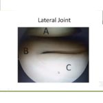
Important Anatomical Landmarks
Key soft tissue landmarks include:
- Posterior horn of the lateral meniscus
- Inverted “J” arch of the femoral footprint
These structures help guide accurate tunnel placement.
Types of ACL Tears and Surgical Implications
Not all ACL tears are identical, and the tear pattern influences graft choice and reconstruction strategy.
1. Mid-Substance Tear
Features:
- No salvageable ACL remnant
- Requires a larger and more robust graft
2. Femoral Detachment
Features:
- Significant remnant preserved
- May require a smaller graft
- Doubled or tripled semitendinosus graft may be sufficient
Understanding the tear type helps tailor graft selection and surgical planning.
Graft Choices in ACL Reconstruction
Patellar Tendon Graft
Historically considered the gold standard during the 1980s and early 1990s.
Advantages:
- Excellent fixation
- Strong graft construct
Disadvantages:
- Higher donor-site morbidity
- Increased anterior knee pain
Hamstring Grafts
Became increasingly popular from the late 1990s onward.
Advantages:
- Reduced donor-site morbidity
- Less anterior knee pain
Quadriceps Tendon Graft
A more recent trend in ACL reconstruction.
Advantages:
- Thick and strong graft
- Better collagen quality
- Larger graft diameter (commonly 9–10 mm)
- Adequate graft length
Modern fixation systems have facilitated the use of quadriceps tendon grafts.
Graft Harvesting Technique
Hamstring Harvest
The pes anserinus is identified first.
Key structures:
- Gracilis tendon lies superiorly
- Semitendinosus tendon is isolated separately
Important technical points:
- Remove all soft tissue attachments before tendon stripping
- Ensure adequate graft length
- Preserve full graft width
Careful harvesting minimizes graft damage and improves graft quality.
Graft Preparation
Common preparation methods include:
- Quadrupled semitendinosus graft
- Tripled semitendinosus graft when additional diameter is required
Synthetic Augmentation
Synthetic augmentation devices such as fiber loops may improve construct strength, but caution is advised because some materials may produce adverse tissue reactions.
Stepwise Surgical Technique
The KISS Principle
The procedure is simplified into a sequential approach:
- Diagnostic arthroscopy
- Graft harvesting
- Tunnel preparation
- Graft passage
- Graft fixation
This systematic approach helps maintain consistency during surgery.
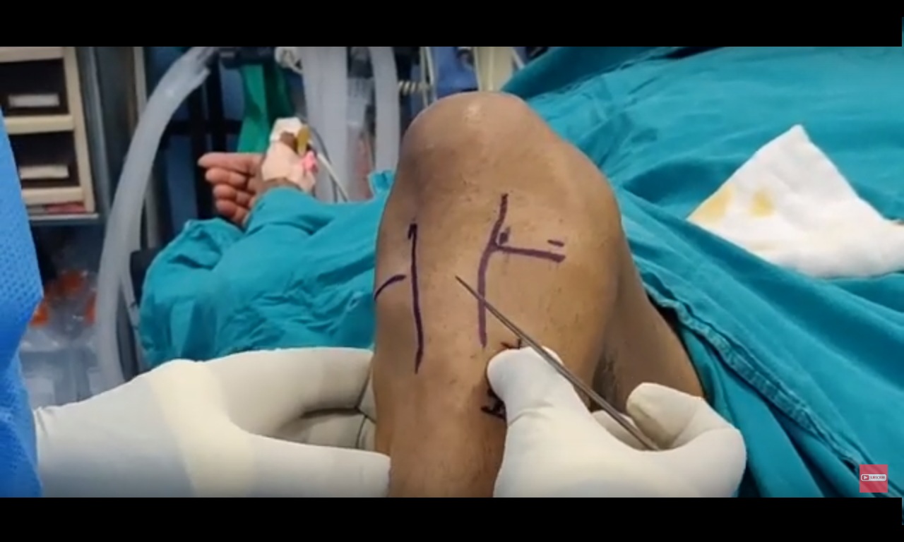
Diagnostic Arthroscopy
A Critical Initial Step
Diagnostic arthroscopy should be performed before graft harvesting.
It helps evaluate:
- ACL tear pattern
- Quality of remnant tissue
- Associated meniscal injuries
- Posterolateral corner injuries
- Medial collateral ligament involvement
This assessment influences:
- Graft selection
- Graft size
- Need for additional procedures
Accurate diagnosis at this stage is essential for proper surgical planning.
Notch Assessment and Notchplasty
When is Notchplasty Needed?
Notchplasty may be necessary in chronic ACL-deficient knees.
Benefits include:
- Improved visualization of the femoral footprint
- Easier tunnel placement
However, unnecessary notchplasty should be avoided to preserve normal anatomy.
Femoral Tunnel Placement
Common Errors
Incorrect tunnel positioning remains one of the major causes of graft failure.
Common mistakes include:
- Tunnel placed too anteriorly
- Tunnel placed too posteriorly
Preferred Technique
The transportal approach is commonly preferred because it allows independent femoral tunnel placement.
Drilling Options
Rigid Jig
Limitations:
- Requires hyperflexion
- Visualization may be difficult
Flexible Reamer
Advantages:
- Knee maintained at approximately 90° flexion
- Better visualization
- Preserves fat pad and synovium
Flexible reaming systems are increasingly favored for anatomical tunnel placement.
Tibial Tunnel Placement
Importance of Tibial Tunnel Position
Although sometimes underestimated, tibial tunnel placement is critical for graft function.
Errors and Consequences
Too anterior:
- Graft impingement
- Stretching and failure
Too posterior:
- Vertical graft orientation
- Poor rotational control
Too medial or lateral:
- Cartilage injury
Important Landmarks
Key landmarks include:
- Medial tibial spine
- Posterior border of the anterior horn of the lateral meniscus
These landmarks help guide accurate tunnel placement.
Tibial Tunnel Technique
Serial Dilatation Technique
The tunnel is gradually enlarged using sequential dilators.
Technique:
- Start with K-wire placement
- Gradually increase tunnel size
Advantages:
- Reduces risk of tibial plateau fracture
- Particularly useful in young athletes with dense bone
Correction of Tunnel Misplacement
If the initial guide pin is malpositioned:
- Leave the original pin in place
- Insert a second pin in the correct position
This prevents accidental reuse of the incorrect tunnel trajectory.
Graft Passage
Principles
The graft should pass smoothly without resistance.
Forceful passage may result in:
- Graft laceration
- Weakening of the graft construct
Tunnel dilators may be used before graft passage to smooth the tunnel and reduce resistance.
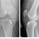
Femoral Fixation
Endobutton Fixation
Key point:
- Ensure the Endobutton flips properly on the lateral femoral cortex
Visualization is usually achieved through:
- Central portal
- Anteromedial portal
Secure fixation is essential for graft stability during healing.
Final Arthroscopic Assessment
Before completing the procedure:
- Remove debris anterior to the graft
- Prevent cyclops lesion formation
- Confirm full extension and flexion
- Ensure there is no graft impingement
Final assessment is essential to optimize postoperative knee function.
The ABCD Concept of ACL Reconstruction
A useful summary principle includes:
A — Anatomic Placement
- Accurate tunnel positioning
B — Biological Preservation
- Preserve ACL remnant tissue
- Preserve soft tissue structures
C — Collagen Quality
- Use a strong and appropriately sized graft
D — Durable Fixation
- Achieve stable graft fixation
Final Surgical Goals
The ultimate goals of ACL reconstruction are:
- Strong graft construct
- Anatomical tunnel placement
- No graft impingement
- Preservation of biology
- Secure fixation
- Identification and treatment of associated injuries
Successful ACL reconstruction depends on combining precise surgical technique with sound biological principles.
Leave a Reply