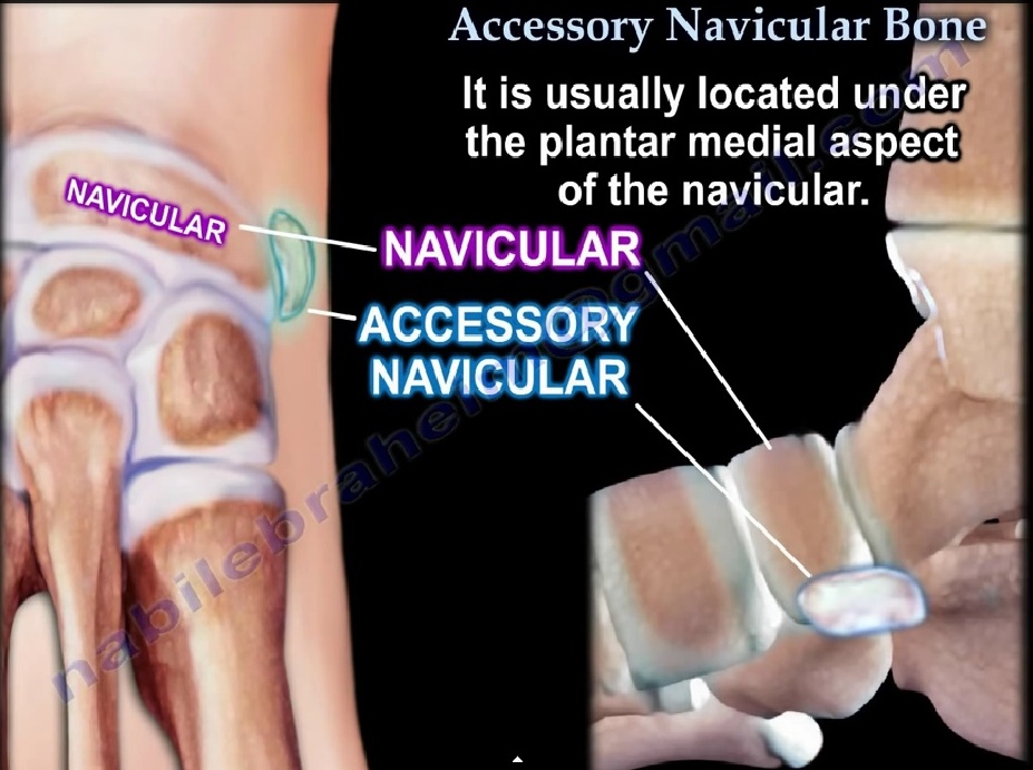
• Skip to main content
  • Skip to secondary menu
  • Skip to primary sidebar
OrthopaedicPrinciples.com

OrthopaedicPrinciples.com

Integrating Principles and Evidence

Integrating Principles and Evidence

  • Home
  • Editorial Board
  • Our Books
    • Evidence Based Orthopaedic Principles
  • Courses
  • Exams
  • Reviews
  • Live Program
  • Contact

Accessory Navicular

Accesory Navicular

  • This anatomic variant consists of an accessory ossicle located at the medial edge of the navicular
  • Accessory ossicles are derived from unfused ossification centers.
  • 90% bilateral
  • It is most commonly symptomatic in the 2nd decade of life and causes medial foot pain
  • Symptomatic in <1% of patients.
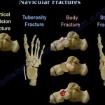
Classification

  • Type I occurs primarily as a round sesamoid within the substances of the distal posterior tibial tendon.

It is rarely associated with symptoms.

Small, 2–3-mm sesamoid bone in the PTT; referred to AS “os tibiale externum”


  • Type II is associated with a synchondrosis within the body of the navicular at risk for disruption either from traction injury or shear forces in the region.

Larger ossicle than type I

Secondary ossification center of the navicular bone

Most common variety (50%)


  • Type III, also known as a navicular beak or a cornuate navicular occurs with fusion of the accessory navicular bone to the body of the navicular.


Clinical Features:

–         Pain may begin after wearing ill-fitting shoes, with weight bearing activities or athletics, or after trauma to the foot.

–         Tenderness over the medial aspect of the foot and over the accessory navicular bone.

–         Secondary Achilles tendon contracture can occur

–         Flatfoot is common and with severe flatfoot, lateral pain may occur secondary to impingement of the calcaneus against the fibula.

X-rays:

–         AP, lateral, internal oblique and external oblique

–         The accessory ossicle may be best visualized on the internal oblique view(reverse oblique view)


Treatment

•   Conservative measures: Shoe-wear modification, including use of a softer, wider shoe,  NSAIDS

•   Medial arch support for flatfoot.

•   A below knee cast may be worn for 3 -6 weeks for persistent symptoms

•   Surgery maybe employed for persistent pain not responding to conservative measures

•    Kidner’s procedure: Consists of excising the accessory navicular and rerouting the tibialis posterior tendon into a more plantar position.

•    Kidner’s operation will not correct sag at the metatarsocuneiform joint.

•   Severe flatfoot deformity with lateral impingement symptoms may require concomitant osteotomy of the calcaneus and/or medial column of the foot to improve alignment and decrease mechanical stress of the PTT insertion.

Ref:

  1. Sella EJ, Lawson JP, Ogden JA. The accessory navicular synchondrosis. Clin Orthop Relat Res 1986;209:280–285

Post Views: 7,289

Related Posts

  • Accessory Navicular

    Courtesy: Prof Nabile Ebraheim, University of Toledo, Ohio, USA

  • Navicular Fractures

    Courtesy:Prof Nabil Ebraheim,University of Toledo, Ohio, USA

  • Evidence Based Medicine

    1. A powerpoint presentation on Evidenced Based Medicine, by one of the pioneer spine surgeons…

Reader Interactions

Leave a Reply

Your email address will not be published. Required fields are marked *

Primary Sidebar

Follow Us

instagram slideshare

Categories

  • -Applied Anatomy
  • -Approaches
  • -Basic Sciences
  • -Cartilage & Meniscus
  • -Classifications
  • -Examination
  • -Foot and Ankle
  • -Foot and Ankle Trauma
  • -FRCS(Tr and Orth) tutorials
  • -Gait
  • -Hand and Wrist
  • -Hand and Wrist Trauma
  • -Hand Infections
  • -Hip and Knee
  • -Hip Preservation
  • -Infections
  • -Joint Reconstruction
  • -Knee Arthroplasty
  • -Knee Preservation
  • -Metabolic Disorders
  • -Oncology
  • -OrthoBiologics
  • -OrthoPlastic
  • -Paediatric Orthopaedics
  • -Paediatric Trauma
  • -Patellofemoral Joint
  • -Pelvis
  • -Peripheral Nerves
  • -Principles
  • -Principles of Surgery
  • -Radiology
  • -Rheumatology
  • -Shoulder and Elbow
  • -Shoulder and Elbow Arthroplasty
  • -Spine Deformity
  • -Spine Oncology
  • -Spine Trauma
  • -Spine, Pelvis & Neurology
  • -Sports Ankle and Foot
  • -Sports Elbow
  • -Sports Knee
  • -Sports Medicine
  • -Sports Medicine Hip
  • -Sports Shoulder
  • -Sports Wrist
  • -Statistics
  • -Technical Tip
  • -Technology in Orth
  • -Trauma
  • -Trauma (Upper Limb)
  • -Trauma Life Support
  • -Trauma Reconstruction
  • Book Shelf
  • Book Shelf Medical
  • Careers
  • Case Studies and Free Papers
  • DNB Ortho
  • Evidence Based Orthopaedic Principles
  • Evidence Based Orthopaedics
  • Exam Corner
  • Fellowships
  • Guest Editor
  • Guest Reviews
  • Image Quiz
  • Instructional Course Lectures
  • Journal Club
  • MCQs
  • Meetings and Courses
  • Multimedia
  • News and Blog
  • Plaster Techniques
  • Podcasts
  • Public Health
  • Rehabilitation
  • Research
  • Shorts and Reels
Copyright@orthopaedicprinciples.com. All right rerserved.