By Peter Sturm MD
Courtesy : Dr Sanjay Chaturvedi, Organising Secretary, IOACON Agra
Definition
-
Early onset scoliosis is defined as any spinal deformity present before the age of 10 years, irrespective of etiology.
-
Earlier onset is associated with:
-
Greater curve progression
-
Worse long-term prognosis
-
-
Children with EOS have a higher risk of developing severe spinal deformity with increased morbidity and mortality.
-
Progressive deformity restricts thoracic growth and volume.
-
This can result in:
-
Thoracic insufficiency syndrome
-
Cardiovascular compromise
-
Respiratory failure
-
Measurement of Spinal Curvature
-
Spinal curvature is measured using the Cobb angle on radiographs.
Severity Based on Cobb Angle
-
Mild: less than 25°
-
Moderate: 25° to 50°
-
Severe: greater than 50°
Etiological Classification of Early Onset Scoliosis
Early onset scoliosis includes the following categories:
-
Infantile idiopathic scoliosis
-
Juvenile idiopathic scoliosis
-
Congenital scoliosis
-
Neuromuscular scoliosis
-
Syndromic scoliosis
-
Scoliosis associated with tumors, infection, prior surgery, or trauma
Infantile Idiopathic Scoliosis
-
Occurs in children aged 3 years or younger.
-
Accounts for approximately 4% of idiopathic scoliosis cases.
-
More common in males than females.
-
Typically presents with a left thoracic curve.
-
Family history is often positive.
Juvenile Idiopathic Scoliosis
-
Occurs between 4 and 10 years of age.
-
Accounts for approximately 15% of idiopathic scoliosis cases.
-
More common in females than males.
-
Most commonly presents as a right main thoracic curve.
Congenital Scoliosis
-
Results from failure of normal vertebral development during the 4th to 6th week of gestation.
-
Incidence ranges from 1% to 4% in the general population.
-
May occur in isolation or in association with other systemic anomalies.
Neuromuscular Scoliosis
-
Caused by disorders of the brain, spinal cord, or muscular system.
-
Curves tend to:
-
Progress rapidly
-
Be long and involve multiple vertebrae
-
Occasionally involve the cervical spine
-
Syndromic Scoliosis
-
Occurs in association with genetic or systemic syndromes, including:
-
Marfan syndrome
-
Prader–Willi syndrome
-
Trisomy 21 (Down syndrome)
-
Ehlers–Danlos syndrome
-
Osteochondrodystrophy (dwarfism)
-
Neurofibromatosis
-
Clinical Presentation
General Presentation
-
Many children appear normal and function well, especially with mild curves.
-
Most present due to visible deformity.
-
Back pain is uncommon.
-
Neurologic symptoms are usually absent.
Physical Examination
Key Principle
-
Careful assessment of body symmetry is essential for detecting scoliosis and progression.
Inspection Findings
-
Uneven or tilted shoulders
-
Prominence of one scapula
-
Spine deviating from the midline
-
Head not centered over the pelvis
-
Rib prominence on one side
-
Unequal hip levels
-
Asymmetric waistline
-
Inability to stand erect
-
Leaning to one side
-
Asymmetric skin creases
-
Pain is uncommon but may occasionally be present
General Examination
-
Café-au-lait spots suggesting neurofibromatosis
-
Cutaneous nevi
-
Limb length discrepancy
-
Signs of neural axis abnormalities
-
Foot deformities such as cavovarus
-
Gait analysis
Spine Examination
-
Midline skin abnormalities:
-
Hairy patches
-
Dimples
-
Signs suggestive of spinal dysraphism
-
-
Rib rotational deformity or rib hump
-
Adam forward bending test:
-
Demonstrates axial plane deformity in structural scoliosis
-
-
Forward bending test in sitting position:
-
Eliminates leg length inequality as a cause
-
-
Plagiocephaly in younger children
Neurologic Examination
-
Assessment of motor development and milestones
-
Evaluation of upper and lower limbs
-
Screening for cavovarus feet
-
Reflex assessment:
-
Abdominal reflexes
-
Clonus
-
Hoffman sign
-
Babinski response
-
Investigations
Radiographs
-
Recommended views:
-
Standing posteroanterior view
-
Standing lateral view
-
-
Evaluate for congenital vertebral anomalies.
Measurements
-
Cobb angle
-
Rib phase
-
Rib vertebral angle difference (Mehta angle)
Magnetic Resonance Imaging
-
Used to identify neural axis abnormalities.
-
Mandatory in all patients with congenital scoliosis prior to surgery.
-
Indicated in:
-
Curves greater than 20°
-
Presence or absence of neurologic symptoms
-
Computed Tomography
-
Used judiciously due to radiation exposure.
-
Three-dimensional computed tomography is useful for detailed assessment of posterior bony anatomy.
Low-Dose EOS Imaging
-
Uses ultra-low radiation to generate three-dimensional models from two planar images.
Additional Investigations
-
Bone scan or dual-energy X-ray absorptiometry when indicated.
-
Renal ultrasound or magnetic resonance angiography for associated renal anomalies.
-
Echocardiography if cardiac involvement is suspected.
Treatment Goals
-
Slow progression of the spinal curve.
-
Correct deformity while allowing spinal and thoracic growth.
-
Maximize pulmonary function.
-
Preserve chest wall and spinal mobility.
-
Consider overall growth and development of the child.
-
Prevent or delay definitive spinal fusion.
-
Minimize complications, procedures, hospitalizations, and family burden.
Conservative Management Options
-
Observation and close monitoring
-
Serial casting
-
Bracing using thoracolumbosacral orthosis
-
Halo gravity traction
Surgical Management Options
-
Non-fusion surgery
-
Definitive spinal fusion surgery
Non-Fusion Surgery
-
Indicated for curves greater than 50° in growing children.
-
Allows continued spinal growth across unfused segments.
-
Definitive fusion is delayed until near skeletal maturity.
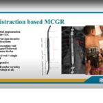
Distraction-Based Systems
-
Correct deformity by applying distractive forces to the spine, ribs, or pelvis.
Examples
-
Traditional growing rods
-
Magnetically controlled growing rods
-
Vertical expandable prosthetic titanium rib system
Compression-Based Implants
-
Modulate growth by applying compression on the convex side of the curve, inhibiting growth.
Examples
-
Vertebral body stapling
-
Vertebral body tethering
Guided Growth Systems
-
Use fixed and non-fixed anchors connected to rods to guide spinal growth.
Examples
-
Luque trolley technique
-
Shilla growth guidance technique
Other Surgical Alternatives
-
Vertebral column resection
-
Convex hemiepiphysiodesis or convex growth arrest
-
Rib osteotomies
Spinal Fusion Surgery
-
Permanently joins 2 or more vertebrae to prevent further deformity progression.
-
Limits motion at fused segments.
Types
-
Anterior spinal fusion
-
Posterior spinal fusion
Prognosis
-
Depends on the risk of curve progression and timing of intervention.
-
Non-progressive curves may resolve spontaneously.
-
Progressive curves have poorer outcomes and require treatment.
Predictors of Progression (Mehta Criteria)
-
Documented progression of Cobb angle greater than 20°
-
Rib vertebral angle difference greater than 20°
-
Phase 2 rib–vertebral relationship with rib overlap
-
Presence of a double major curve pattern
Crankshaft Phenomenon
-
Refers to loss of three-dimensional correction after spinal surgery.
-
Occurs due to continued anterior spinal growth following posterior fusion in skeletally immature patients.
Leave a Reply