
Courtesy: Dr Taylor J Reif, Hospital for Special Surgery New York
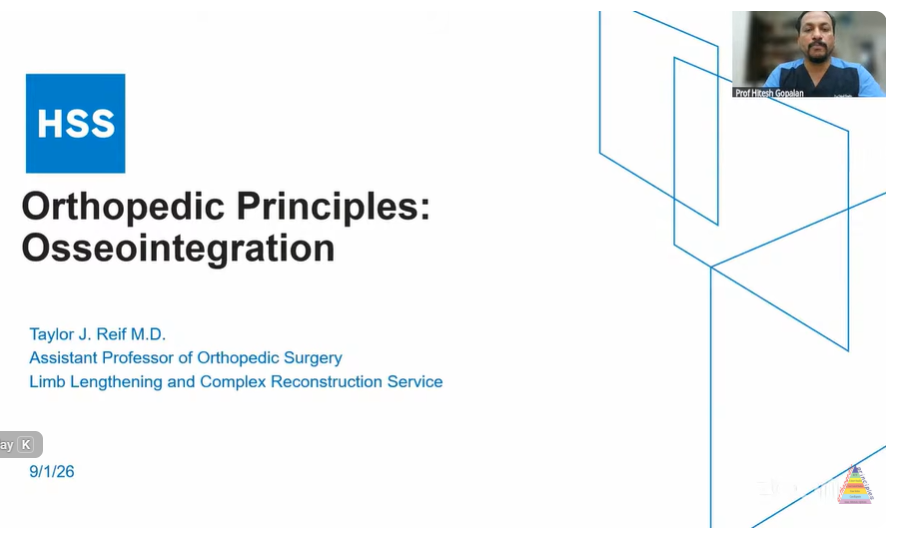
Osseointegration in Amputee Reconstruction: Indications, Implantation, and Soft-Tissue Principles
1. Limitations of Conventional Socket Prostheses
-
Socket-Interface Biomechanics:
-
Conventional sockets act as an uncoupled interface; cyclical pistoning and dynamic displacement of the residual bone inside the soft-tissue sleeve alter mechanical load transfer.
-
Suspended residual limbs lack direct skeletal alignment, often leading to persistent abductor lurch, compensatory gait mechanics, and energy expenditure spikes.
-
-
Cutaneous and Soft-Tissue Morbidity:
-
High prevalence of friction ulcers, shear blister formation, pressure necrosis, allergic contact dermatitis, and recurrent superficial/deep infections.
-
Heterotopic ossification, angular bony prominences, muscle atrophy, cutaneous grafts, and fluctuating limb volume (edema, perspiration, temperature swings) frequently prevent consistent socket fit.
-
-
Neuroma and Phantom Pain:
-
Transected peripheral nerves form traction/terminal neuromas (5–15\% or more of residual limbs), causing focal hypersensitivity exacerbated by external socket wall compression.
-
-
Prosthetic Abandonment:
-
High attrition rate: up to 40–50% of transfemoral amputees struggle with functional ambulation, frequently abandoning prostheses or requiring annual socket refabrications.
-
2. Evolution, Implant Biomechanics, and Fixation Concepts
-
Osseointegration Principles:
-
First coined by Brånemark in 1977 based on direct bone-to-titanium contact without an intervening fibrous tissue layer.
-
Direct structural anchoring restores the patient’s anatomical mechanical axis and provides “osseoperception” (enhanced vibratory feedback through the bone).
-
-
Implant Designs & Fixation Mechanics:
-
Threaded Screw-In Systems (OPRA – Sweden):
-
Two-stage procedure: intramedullary threaded fixture (approx. 80 mm length) inserted and bone-grafted (Stage 1), followed by a 3- to 6-month biological latency before inserting the transcutaneous abutment (Stage 2).
-
-
Press-Fit Coated Stems (Integral Accompanying Prosthesis [ILP]/Bad Oeynhausen – Germany; Osseointegrated Prosthetic Limb [OPL] – Australia):
-
Single- or two-stage insertion using textured/porous proximal coatings for biological ingrowth, coupled to a smooth distal transcutaneous neck (titanium-niobium nitride) to prevent cutaneous abrasion.
-
Modular attachment mechanisms (dual cones, taper sleeves, bushings) secure the external exoprosthesis.
-
-
Custom & Compliant Loading Systems:
-
Electron-beam melted (EBM 3D-printed) custom titanium constructs for ultra-short residual limbs.
-
Adaptations of compressive osseointegration devices (e.g., Compress technology) utilizing spring-loaded cortical spindles for compliant biological interface loading.
-
-
-
Aseptic Loosening vs. Stress Risers:
-
CT-based 3D modeling and tight canal fill minimize interface micromotion; true aseptic loosening is rare.
-
Rigid intramedullary fixation creates a proximal stress riser at the host bone-implant junction, predisposing to periprosthetic fractures (5–10%) following falls.
-
3. Surgical Technique & Crucial Soft-Tissue Principles
-
Bone Preparation:
-
Canal reaming and broaching matched precisely to preoperative CT dimensions to prevent uncoupled gaps and fibrous encapsulation.
-
In active osteomyelitis or compromised residual bone stock, a two-stage approach (bone debridement, antibiotic spacer, and clearance interval) is mandatory prior to hardware placement.
-
-
The “Soft-Tissue Paradigm”:
-
Surgical Axiom: The osseous reconstruction is predictable; soft-tissue aperture/stoma management represents the principal clinical challenge.
-
Elimination of Deep Adipose: Aggressive debulking of deep subcutaneous fat down to the dermis, creating a coned-down distal stump.
-
Myoplasty / Muscle Collar: Avoid distal muscular overhang around the implant neck. Muscle is sharply retracted or trimmed back to the level of the cortical bone and anchored as a tight proximal cuff (“fascial sock”).
-
Aperture Stability (Skin-to-Bone Adherence): The dermis is quilted directly to the periosteum/bone cortex with tension-free margins. Motion and shear at the skin-implant junction are the primary etiologies of stoma irritation, granulation tissue formation, and ascending infection.
-
Tibia-Specific Advantage: Transtibial segments feature a thinner native soft-tissue envelope, facilitating rigid skin adherence and eliminating crossing-joint socket impingement.
-
-
Peripheral Nerve Management:
-
Concomitant execution of Targeted Muscle Reinnervation (TMR) or Regenerative Peripheral Nerve Interfaces (RPNI) using autologous muscle grafts to cap transected nerves, preventing symptomatic neuromas and mitigating phantom limb pain.
-
4. Postoperative Rehabilitation & Staged Loading Protocol
-
Quiescence Interval: 2 to 4 weeks of limb rest postoperatively to permit surgical soft-tissue settling and initial stoma sealing.
-
Progressive Loading Phase (“Rubber Footy” Protocol):
-
Application of a distal rubber bumper/stopper directly onto the external abutment.
-
Static axial loading on a floor scale starting at 20 lbs multiple times daily, advancing in 5–10lbs increments every few days up to 100 lbs(or half body weight).
-
Stimulates cortical hypertrophy and bone remodelling prior to full prosthetic ambulation.
-
-
Full Prosthetic Ambulation: Advanced gradually with specialized physical therapy focused on abductor/gluteal re-education to correct ingrained Trendelenburg gait mechanics.
5. Complications, Outcomes, and Survivorship
-
Implant Survivorship: Long-term multicenter data (up to 15 years) demonstrate hardware retention rates exceeding 90–95%; complete explantation occurs in <5% of cases.
-
Skin-Implant Interface Infections:
-
Superficial aperture drainage is frequent, reflecting colonization of an open tract rather than deep joint infection.
-
Managed with local wound hygiene, oral antimicrobials, or minor bedside packing; typically resolves without hardware sacrifice.
-
-
Reoperation Drivers:
-
Soft-tissue revision (redundant tissue excision, thinning of recurrent overhang, aperture release) represents the most common cause for return to the operating room.
-
Periprosthetic fractures proximal to the stem tip are managed with standard open reduction and internal fixation (ORIF) without requiring implant removal.
-
-
Functional and Quality-of-Life Metrics:
-
Patients demonstrate significant improvements in physical function, mobility, PROMIS scores, and two-minute walk tests (>100feet gain).
-
Prosthetic daily wear times regularly reach 14 to 16 hours/day.
-
Particularly impactful in short residual limbs (femoral remnants <8 cm} or tibial segments 4-5 cm) and bilateral amputees otherwise confined to wheelchairs.
-
Master Revision Summary
-
Surgical Concept: Direct skeletal attachment of exoprostheses via an osseointegrated titanium stem, eliminating socket-related friction, ulcers, and unstable biomechanical lever arms.
-
Implant Classes: Threaded two-stage fixtures (OPRA) vs. single/two-stage press-fit stems (ILP, OPL) featuring textured bone-anchoring proximal zones and ultra-smooth transcutaneous necks.
-
Critical Technical Step: Aggressive subcutaneous defatting, proximal muscular resection (“fascial sock”), and direct dermal-to-cortical quilting; abolishing skin shear at the aperture is the key to preventing ascending infection.
-
Nerve Handling: Execution of RPNI or TMR to prevent terminal neuroma formation and mitigate phantom limb pain.
-
Rehabilitation Milestone: 2- to 4-week quiet phase – progressive axial loading using a distal rubber stopper on a weighing scale – gradual transition to full weight-bearing exoprosthetics.
-
Complication Profile: Soft-tissue overhang revision is the most common secondary surgery; periprosthetic fractures (5–10%) occur proximal to the stem tip and are treated with ORIF; aseptic loosening is exceptionally rare; long-term implant survivorship exceeds 90–95%

Leave a Reply