
Courtesy: Dr. V Salil Upasani, Dr Ashok Shyam, Ortho TV
3D Printing in Complex Hip Preservation
Overview
- Three-dimensional printing is emerging as a useful adjunct in orthopaedic surgery, particularly for complex hip deformities and hip preservation procedures.
- Its greatest value is as a complementary technology that supports, rather than replaces, clinical judgment, surgical expertise, and conventional imaging.
- Patient-specific three-dimensional models can be used for:
- Preoperative planning.
- Understanding complex three-dimensional deformities.
- Surgical simulation.
- Patient and family education.
- Resident and fellow training.
- Orthopaedic research.
- Development and testing of patient-specific implants and instruments.
Applications in Orthopaedic Surgery
- Three-dimensional printing has been investigated for several challenging orthopaedic procedures.
- Reported applications include:
- Planning correction of congenital spinal deformities.
- Surgical planning for complex pelvic bone tumors.
- Patient-specific distal radius osteotomy planning.
- Complex proximal femoral reconstruction.
- Pediatric pelvic osteotomy planning.
- Simulation and training for technically demanding procedures.
Patient-Specific Models for Surgical Planning
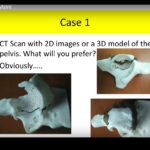
- A three-dimensional model can convert conventional computed tomography data into a physical representation of the patient’s anatomy.
- This allows the surgeon to:
- Directly visualize deformity from multiple perspectives.
- Identify the location and extent of abnormal bone morphology.
- Simulate different osteotomy options.
- Determine the likely amount and direction of correction.
- Assess implant positioning before surgery.
- Anticipate technical difficulties during the procedure.
- A mock operation performed on the printed model can provide a practical rehearsal before undertaking the actual operation.
Application in Slipped Capital Femoral Epiphysis
- Slipped capital femoral epiphysis produces a complex three-dimensional deformity of the proximal femur.
- Severe deformity may require a corrective proximal femoral osteotomy.
- Three-dimensional models can assist in understanding the individual components of the deformity and planning the desired correction.
Three-Plane Proximal Femoral Osteotomy
- A three-plane corrective osteotomy can address the deformity through controlled correction in multiple planes.
- Patient-specific models can be used before surgery to:
- Determine the osteotomy configuration.
- Simulate the correction.
- Estimate the amount of bone resection.
- Plan implant placement.
- Anticipate the final alignment.
Reported Benefits
- In a single-institution clinical study, patients treated using three-dimensional models were compared with patients treated without models and with a historical cohort.
- The use of patient-specific models was associated with:
- Reduced operative time.
- Reduced fluoroscopy exposure.
- Comparable radiographic correction.
- There was no significant difference in blood loss.
- The important finding was not necessarily that three-dimensional planning produced a superior final correction, but that similar correction could be achieved more efficiently.
- The study also reported fewer implant-related technical problems in the model-assisted group.
Limitations
- The findings should be interpreted cautiously because:
- The sample size was small.
- The study was not a prospective randomized comparison.
- The groups had different characteristics.
- Follow-up was not uniform.
- Larger prospective studies are required before definitive conclusions can be drawn regarding the clinical superiority of three-dimensional printing.
Three-Dimensional Planning of Pediatric Pelvic Osteotomies
- Acetabular dysplasia requires individualized correction because the pattern of acetabular deficiency varies between patients.
- Common pelvic osteotomies used in children include:
- Dega osteotomy.
- Pemberton osteotomy.
- San Diego osteotomy.
- Although these procedures may appear similar externally, their osteotomy paths and mechanisms of correction differ substantially.
Understanding the Osteotomy Geometry
- Three-dimensional printed pelvic models allow the surgeon to appreciate the internal anatomy and the relationship between:
- The osteotomy.
- The acetabulum.
- The triradiate cartilage.
- The medial wall of the ilium.
- The posterior pelvic structures.
- The different osteotomies produce different patterns of acetabular reorientation and coverage.
Simulation Using Identical Patient-Specific Pelvic Models
- A particularly useful research approach is to produce multiple models of the same patient’s pelvis.
- Different osteotomies can then be performed on separate copies of the model.
- Three-dimensional computed tomography analysis can subsequently be used to compare the resulting changes in acetabular coverage.
Principal Findings
- Different pelvic osteotomies produce different patterns of acetabular coverage.
- In the reported experimental analysis:
- Pemberton and Dega osteotomies produced greater anterior coverage.
- San Diego osteotomy produced relatively greater posterior coverage.
- This supports the principle that pelvic osteotomy should be selected according to the individual pattern of acetabular deficiency, rather than applying a single technique uniformly to every patient.
- The models also demonstrated a reduction in acetabular volume following correction.
- The reduction in volume was broadly comparable among the osteotomy techniques evaluated.
Complex Proximal Femoral Reconstruction After Infection
- Three-dimensional printing can be particularly valuable when conventional imaging makes it difficult to appreciate the complete extent of a deformity.
- An example involved a young child with:
- Septic arthritis of the hip.
- Femoral osteomyelitis.
- Extensive surrounding muscle inflammation.
- Methicillin-resistant Staphylococcus aureus infection.
- The infection required repeated surgical debridement and prolonged antimicrobial treatment.
- Following control of the infection, the child developed:
- A pathological fracture.
- Extensive destruction of the femoral canal.
- Malunion.
- Proximal femoral deformity.
- Varus deformity at the femoral head-neck region.
- Rotational deformity.
- Associated shaft deformity.
Role of Three-Dimensional Printing
- A patient-specific three-dimensional model of the pelvis and both proximal femora was produced.
- The model enabled detailed assessment of:
- The residual deformity.
- The relationship between the femoral head, neck, and shaft.
- Possible osteotomy sites.
- The feasibility of reconstruction.
- Implant selection and modification could also be tested on the model before surgery.
- A small plate was customized to fit the patient’s unusually small and deformed proximal femur.
- After appropriate sterilization and surgical preparation, the customized implant was used during reconstruction.
- Follow-up demonstrated substantial improvement in proximal femoral alignment and biomechanics after reconstruction and subsequent implant removal.
Complex Residual Perthes-Type Proximal Femoral Deformity
- Another application involved an adolescent with a complex proximal femoral deformity following previous hip disease consistent with residual Perthes-type deformity.
- Three-dimensional computed tomography demonstrated abnormalities that were difficult to appreciate fully on conventional two-dimensional radiographs.
Surgical Options Considered
- Several reconstructive procedures were evaluated on the patient-specific model, including:
- Valgus femoral osteotomy.
- Femoral neck lengthening procedures.
- Intertrochanteric osteotomy.
- Surgical dislocation with femoral head reduction osteotomy.
- Performing these procedures on a physical model allowed direct comparison of the expected anatomical correction.
Patient-Specific Surgical Selection
- After simulation, a multi-part proximal femoral corrective osteotomy was selected.
- Postoperative imaging demonstrated substantial improvement in:
- Proximal femoral morphology.
- Mechanical alignment.
- Hip biomechanics.
- Serial follow-up showed maintenance of the correction after healing and subsequent implant removal.
Advantages of Three-Dimensional Printing
- Improved anatomical understanding
- Provides a tangible representation of complex deformity.
- Allows visualization of anatomy from multiple angles.
- Better preoperative planning
- Helps determine osteotomy location and orientation.
- Allows simulation of correction before surgery.
- Facilitates implant planning.
- Surgical rehearsal
- Complex procedures can be practiced before entering the operating room.
- Potential technical problems can be identified in advance.
- Potential reduction in operative time
- Preoperative simulation may improve surgical efficiency.
- Reduced radiation exposure
- Better preoperative planning may reduce dependence on intraoperative fluoroscopy in selected procedures.
- Education and training
- Provides residents and fellows with realistic models for practicing complex procedures.
- Allows repeated simulation without exposing patients to operative risk.
- Patient and family communication
- Physical models can make complex deformities easier for patients and families to understand.
- Research
- Identical patient-specific models can be used to compare different surgical techniques under controlled conditions.
- Patient-specific implants
- Customized implants or guides can potentially be designed for unusual anatomy.
Practical Considerations
- Three-dimensional printing does not replace:
- Clinical examination.
- Conventional radiography.
- Computed tomography.
- Magnetic resonance imaging when indicated.
- Sound surgical judgment.
- Its value depends on accurate imaging data, appropriate segmentation, reliable model production, and careful surgical interpretation.
- The technology is particularly useful when the deformity is:
- Complex.
- Three-dimensional.
- Unusual.
- Difficult to understand using conventional imaging alone.
Cost and Accessibility
- Desktop three-dimensional printers can provide a relatively accessible option for producing anatomical models.
- The cost of the printer may be modest compared with many advanced medical technologies, while the material cost of an individual model can be relatively low.
- Actual clinical implementation also requires consideration of:
- Imaging and segmentation software.
- Personnel and technical expertise.
- Model production time.
- Sterilization requirements when models or customized implants are taken into the operating room.
- Regulatory and quality-control requirements for patient-specific implants.
Key Takeaways
- Three-dimensional printing is a supportive surgical planning technology, not a substitute for surgical expertise.
- Patient-specific models are particularly valuable in complex hip and proximal femoral deformities.
- Physical simulation can improve understanding of deformity and allow comparison of different reconstructive strategies before surgery.
- Different pelvic osteotomies produce different patterns of acetabular correction, emphasizing the importance of individualized surgical planning.
- Three-dimensional printing has potential applications in:
- Preoperative planning.
- Surgical simulation.
- Resident education.
- Research.
- Patient communication.
- Customized implant development.
- Current evidence is promising but remains limited by small study populations and heterogeneous study designs.
- The greatest potential benefit lies in combining advanced technology with sound clinical judgment and surgical skill.



Leave a Reply