
Courtesy: Ms. Ruth Delaney, MB BCh, MMedSc, FFSEM, FRCS
Founder, Dublin Shoulder Institute
Consultant Orthopaedic Surgeon, Shoulder Surgery Specialist, Associate Professor
The Rationale for 3D Preoperative Planning
-
The Longevity Problem: Glenoid component failure remains the primary mode of arthroplasty breakdown, especially in young arthritic patients.
-
Limitations of Unassisted Experience: Relying on unassisted experience reproduces the wide variability and errors of historic surgery.
-
The Retroversion Penalty: In anatomic total shoulder arthroplasty (aTSA), implanting a glenoid component in 15 degrees of retroversion versus neutral causes asymmetric edge loading, focal contact stress, and early aseptic loosening.
-
The Superior Tilt Penalty: In aTSA, superior component tilt directly causes secondary rotator cuff failure. In reverse shoulder arthroplasty (RSA), superior baseplate tilt causes early mechanical shear and catastrophic failure.
-
The RSA Angle (Pascal Boileau Concept): The inferior glenoid face often displays greater superior inclination than the global glenoid cavity. Planning must orient the baseplate relative to this local inferior angle to prevent inadvertent superior tilt.
Diagnostic Imaging Modality Comparisons
-
Plain Radiographs:
-
Uses true AP (Grashey) and axillary lateral views.
-
Shows gross bony anatomy only.
-
Prone to severe overestimation or underestimation of version and tilt.
-
-
2D CT Scans:
-
Uses standard axial and coronal reconstructions.
-
Relies on manual multi-slice measurements such as the Friedman line.
-
Associated with high inter-observer error and inconsistent landmark selection.
-
-
3D CT Planning Software:
-
Uses complete volumetric scapular segmentation.
-
Provides automated multi-point surface mapping.
-
Can occasionally be fooled by large peripheral osteophytes.
-
3D CT Protocols and Software Segmentation
-
CT Acquisition Protocol: Thin-slice acquisition (1.0 to 1.25 mm cuts) covering the entire body of the scapula down to the inferior angle.
-
Manual vs. Automated Segmentation:
-
Manual segmentation requires engineers to trace individual DICOM slices, introducing a 24 to 48 hour delay.
-
Automated algorithms segment DICOM volumes in minutes, enabling immediate point-of-care surgical planning.
-
-
Software Accuracy: Software measurements for version and inclination are accurate and accepted without revision over 90 to 95 percent of the time.
-
Software Limitations: Large peripheral osteophytes can distort automated surface landmarks. Severe deformities (such as Walch Type C glenoids with over 50 degrees of retroversion) produce divergent measurements across commercial planning platforms.
Modern Guidance Modalities: From Planning to Execution
-
Patient-Specific Instrumentation (PSI):
-
Custom 3D-printed polyamide guides engineered from the virtual preoperative plan.
-
The guide feet register directly onto the un-reamed glenoid face to replicate pin trajectory, version, and tilt.
-
Never excise peripheral glenoid osteophytes before applying the PSI guide; removing osteophytes destabilizes planned footplate seating.
-
Production and sterilization lead times range from 2 weeks in the United States to 4 weeks in Europe and Ireland.
-
-
Computer-Assisted Navigation: Optical arrays provide intraoperative feedback without manufacturing delays, but require capital equipment.
-
Robotic-Assisted Arthroplasty: Emerging tool designed to execute difficult, restricted glenoid bone preparation without direct line-of-sight access.
-
Virtual Range-of-Motion (ROM) Simulation: Software predicts impingement-free arcs of motion. These models treat the scapula as rigid, ignore dynamic scapulothoracic motion, and cannot account for deltoid or neurovascular soft-tissue tension.
Mixed Reality (MR) via Microsoft HoloLens 2
-
Virtual Reality (VR): Completely synthetic digital environment used for gaming, education, and simulation.
-
Augmented Reality (AR): Static 2D or 3D digital overlays placed onto the physical environment.
-
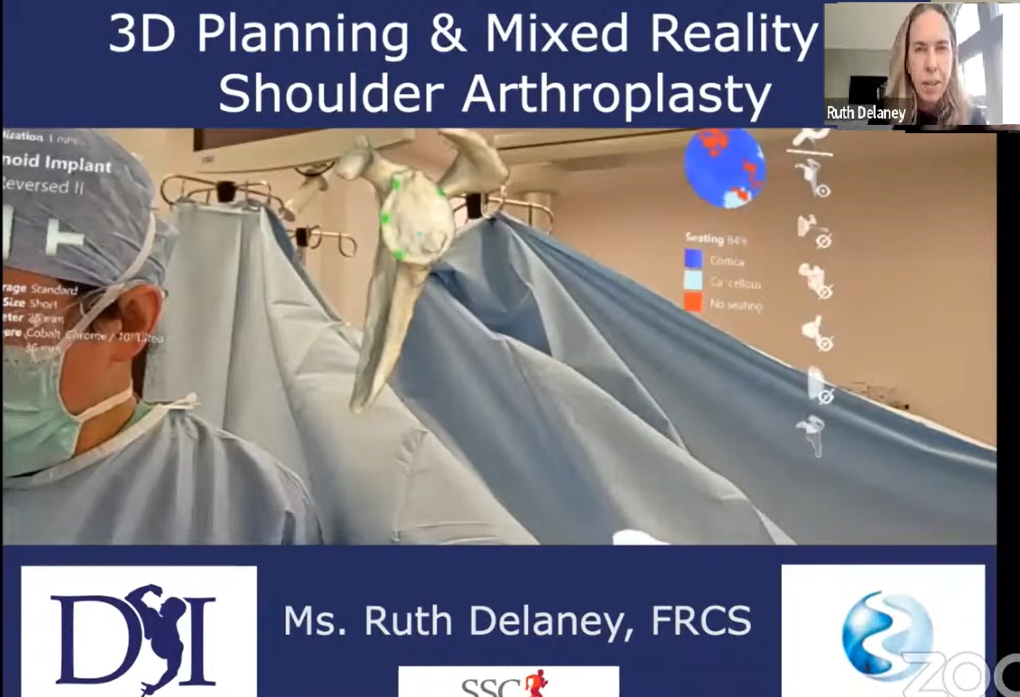
Mixed Reality (MR): Interactive, dynamic holograms anchored to real operative anatomy that the user can manipulate in real time.
-
Operative Usability: Lightweight, untethered head-mounted holographic computer that avoids the large capital footprint of navigation towers or surgical robots.
-
Visual Optics: Visor features a faint tint resembling light sunglasses. Overhead operating lights can be dimmed slightly to enhance holographic contrast.
-
Dynamic Holographic Clone:
-
Projects the patient’s segmented 3D bone model directly beside or over the open wound.
-
The surgeon can rotate, slice, resize, and translate the virtual model in space using sterile, touch-free hand gestures.
-
Unconstrained view eliminates the need to break scrub, step away from the patient, or check wall-mounted displays.
-
-
Interactive Multi-View Streaming: The holographic feed streams wirelessly to operating room monitors, providing assistants, trainees, and implant specialists with the primary surgeon’s exact perspective.
Complex Case 1: Severe Glenoid Erosion in Juvenile Rheumatoid Arthritis
-
Demographics: 30-year-old female with juvenile rheumatoid arthritis (JRA).
-
Pathoanatomy: Severe medial glenoid vault erosion extending directly to the base of the coracoid process, leaving virtually no native vault bone stock.
-
Challenge: Young chronological age, exhausted conservative care, severe functional impairment, and poor proximal humeral bone quality.
-
Software Navigation: 3D planning mapped the only viable central bony corridor down the deep scapular vault to anchor the baseplate central post.
-
Bone Graft Strategy: Selected structural bone grafting (BIO-RSA technique) over metal augments to reconstitute the deficient glenoid vault for future revisions.
-
Graft Sourcing: Native humeral bone was structurally osteopenic; an allograft femoral head was substituted.
-
Staged Reconstruction:
-
Stage 1: Structural allograft bone block, reverse baseplate, and glenosphere placed without a humeral implant.
-
Stage 2: Humeral stem placed after 4 months following CT confirmation of complete bone graft incorporation.
-
Mechanical Rationale: Immediate complete reconstruction creates large shear loads across the graft-glenoid junction, risking acute construct avulsion.
-
Complex Case 2: Osteoarthritis with High Retroversion Managed with Mixed Reality
-
Demographics: Active male masters rower and golfer with severe bilateral end-stage osteoarthritis and high retroversion.
-
Logistical Problem: CT scan completed too close to surgery to produce a 3D-printed PSI guide within the 4-week regional production window.
-
Operative Execution: Managed using the Microsoft HoloLens mixed reality system with the Blueprint holographic planning suite.
-
Humerus Preparation: Virtual humeral head clone guided the neck osteotomy, accounting for shaft-neck angle offsets (155 degree cut versus 135 degree stem).
-
Glenoid Pin Navigation: The 3D glenoid hologram was overlaid directly across the exposed articular surface, aligning the manual guide pin to match the planned version and inferior tilt.
-
Reaming Control: Holographic indicators displayed the precise planned subchondral reaming depth, preventing vault perforation and bone loss.
-
Optimized Screw Trajectories: Viewing the complete 3D scapular morphology in the holographic display allowed the surgeon to place longer peripheral locking screws into dense pillar bone.
Structural Bone Grafting (BIO-RSA) vs. Metallic Augments
-
Structural Bone Grafting (BIO-RSA):
-
Restores native bone stock via biological incorporation.
-
Leaves reconstituted bone stock for future revisions.
-
Autograft is free; allografts are inexpensive.
-
Demonstrates equivalent migration to metal on radiostereometric analysis.
-
Carries a potential risk of non-union or resorption (higher in allografts).
-
-
Metallic Baseplate Augments:
-
Non-biologic permanent metallic foreign body.
-
Revision leaves a massive residual bone void.
-
Involves high manufacturing and hardware expense.
-
Demonstrates low migration with dependable initial press-fit stability.
-
Immune to biological resorption.
-
High-Yield Clinical Pearls
-
Never rely on plain radiographs to quantify glenoid version or tilt; thin-slice 3D CT spanning the full scapular body is the definitive standard.
-
The RSA angle often reveals greater local inferior tilt than global glenoid inclination; plan to this inferior zone to avoid superior baseplate failure.
-
Retain peripheral osteophytes during exposure when using PSI guides; removing them alters the bone surface and destabilizes guide seating.
-
Mixed reality provides dynamic, touchless 3D guidance in the sterile field, bypassing the manufacturing delays of patient-specific instrumentation guides.
-
Structural bone grafting (BIO-RSA) restores the glenoid vault in young arthroplasty candidates, preserving bone stock for anticipated future revisions.



Leave a Reply