PIGMENTED VILLONODULAR SYNOVITIS (PVNS)
Excerpts from “Orthopaedic Principles- A Review”
© Hitesh Gopalan U
Definition:
– Pigmented villonodular synovitis (PVNS) is a slow growing lesion of uncertain etiology arising from the synovial membrane, characterized by villous and nodular overgrowths of the synovial membrane of the bursa or the tendon sheath.
– The appendicular skeleton, especially large joints such as the knee and hip joints are frequently involved.
– Synonyms: Until Jaffe in 1941 proposed the term pigmented villonodular synovitis this condition has been known as synovial xanthoma, synovial endothelioma/ fibroendothelioma, Benign fibrous histiocytoma, xanthomatous GCT, Myeloplaxoma, fibrohemosideric sarcoma , Sarcoma fusigigantocellulare.
History:
• 1852: 1st described as neoplastic process due to unrelenting growth pattern, by Chassaignac, eroding surrounding bone and joint tissue, & high recurrence rate post-resection.
• 1865: Simon described a focal form of PVNS
• 1909: Moser described a diffuse PVNS
• 1941: 1st reported & coined by Jaffe et al. as synovitis, shifting from neoplastic to inflammatory foci.
Prevalance:
• Age: 3rd-4th decades of life, rare in children
• Sex: no sex based predilection
• Incidence: 1.8 per million population
• no predilection for any laterality
Etiopathogenesis:
• repetitive trauma (50%) causing recurrent local hemorrhage to affected joint (cf:hemophilics show progressive erosive arthropathies).
• proliferation of the synovium of joints, tendon sheaths or bursae.
• It is a reactive condition, and not a true neoplasm.
• PVNS classically presents as a monoarticular disease, mimicking arthritis.
• Recurrent atraumatic haemarthrosis is a characteristic feature.
• Often aggressive, with marked extra-articular extension.
Types:
Monoarticular involvement (most common), occurs in two forms: localized and diffuse.
Two variants as described by Granowitz –
a. Localized form (LPVNS): focal involvement of the synovium
– Nodular / Sessile or Pedunculated masses.
– Hands & feet
b. Diffuse form (DPVNS) (more common): affects virtually the entire synovium, eg.
– Intra-articular PVNS tends to be of the diffuse form.
– Tendon sheath PVNS (Giant cell tumour of tendon sheath[GCCTS]), the nodular form.
Sites:
• MC site: knee joint, followed by the hip and shoulder.
• Knee:
– anterior compartment common
– mostly at meniscocapsular junction
– synovium in the region of the anterior horn of the medial meniscus is the most common site
– infrapatellar fat pad, suprapatellar pouch, intercondylar notch, anterior horn of the lateral meniscus, and the medial and lateral recesses of the knee have been reported.
• Uncommon : elbow, ankle, shoulder, foot, wrist
• Rare : spine, cervical involvement commoner than thoracic and lumbar
Clinical features:
o Pain (80%)
o Swelling(76%)
o Reduced range of movement(52%)
o Locking(16%)
o Instability/palpable mass(12%)
Type specific features:
• LPVNS:
– if untreated, causes continuous pain and discomfort, limiting ADLs
– at knee often present with signs and symptoms of meniscal pathology (locking, catching, and instability)
– episodic character of joint effusion—the patient may have completely symptom-free periods between exacerbations
• DPVNS:
– Slow, insidious onset of pain, swelling, and
– stiffness in the involved joint
– most or all joints involved
– swelling and pain more pronounced
– decreased range of motion of the affected joint
– poorly localized
– with sometimes extra-articular extension, either primary or recurrent.
– may encroach on major neurovascular structures.
– Osteoarthritis- continued inflammation and joint erosions lead to articular cartilage destruction, may finally need total joint arthroplasty.
Investigation:
Aspiration of joint: characteristically reveals a blood tinged brownish-stained aspirate.
X-ray:
• Soft tissue swelling will be marked due to haemorrhage and lobulated synovial tissue.
• May reveal cysts or erosions in the joint mimicking gout.
• Bony erosions are usually from without, especially in the hip
• periarticular erosions, with a thin rim of reactive bone
• Osteoporosis is characteristically absent
• Can affect the epiphysis
• Reciprocal bony lesions on opposite sides of the joint, despite articular preservation, are highly suggestive of PVNS
• Late feature of joint space narrowing indicates articular cartilage loss, is difficult to distinguish from primary OA.
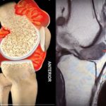
MRI:
• ideal investigation
• nodular mass (periarticular or synovial) with bone erosion
• MRI is invaluable in early diagnosis and evaluating extent. Nodular synovial masses -low signal on T1/T2 sequences
• “dark on dark” on T1- and T2-weighted images
Sonography:
• Loculated joint effusions, Complex heterogeneous echogenic masses and markedly thickened synovium
Arthroscopy:
• direct visualisation of synovium
• Has both diagnostic and therapeutic value in resection of tumours
• Normal arthroscopic findings however does not exclude PVNS (Klompmaker et al)
Histolopathology:
• Synovium looks like a “shaggy carpet”.
• LPVNS is pedunculated, lobular lesion localized to one area of the synovium.
• On microscopy, Histiocytes, lipid laden macrophages, hemosiderin containing cells and frequent giant cells are seen.
• Subsynovial nodular proliferation of large round, polyhedral or spindle cells with prominent cytoplasm and pale nuclei.
Differential diagnosis
• Hemophiliac lobular synovitis (?hemosiderin deposition, lacks lipidladen histiocytes and giant cells, which is classic indications of PVNS)
• Osteoarthritis
• Rheumatoid arthritis,
• Meniscal tear, or other ligamentous injury
Treatment:
• Synovectomy:
o Total synovectomy (open or arthroscopic):
– Open (anterior approach midline incision or medial parapatellar arthrotomy) for the diffuse form for the intraarticular component
– Arthroscopic synovectomy, has gained popularity, has several advantages over the open technique, preferred for LPVNS, shows higher recurrence in DPVNS.
– The standard anterior portals are not effective, whereas the accessory posterior portals are necessary
to accomplish total posterior synovectomy
o Vascular or neurologic injury may occur during this procedure, especially if there is posterior extra-articular
extension of the lesion or fibrosis after irradiation. Open synovectomy should be preferred in such cases
o Open posterior synovectomy (“lazy S-shaped” incision): done subsequently for extensions into the popliteal fossa.
• Local excision: for the nodular form (recurrence rare).
• Radiotherapy (3500- 4000 cGy) (Radiation induced synovectomy/ intra-articular radiation synovectomy using yttrium Y-90) has been used in the management of recurrences with varying success; side effect is soft tissue radionecrosis
• Advanced cases with secondary arthritis should be addressed with arthroplasty plus extensive synovectomy to decrease recurrence.
Prognosis:
• LPVNS: excellent prognosis, low recurrence rate if managed surgically, recurrence 8%.
• DPVNS: surgical excision difficult, recurrence rate of up to 46%.
• The debate continutes: malignant or inflammatory-
– Rare reports describe malignant transformation and metastasis, (presence of trisomy 7 and clonal DNA rearrangements reported).
– Bertoni et al reported eight patients with malignant PVNS; mortality rate was 50%.
– Oehler et al found strong support for its being a chronic inflammatory process and not noeplastic.
– Currently, data are inconclusive to prove PVNS as either malignant or inflammatory process.
– It shows neither cellular atypia nor abnormal mitosis, recent cytogenetic studies say that pathogenesis remains unresolved.
References:
1. Jaffe HL, Lichtenstein L, Sutro CJ. Pigmented villonodular synovitis, bursitis and tenosynovitis. Arch Pathol 1941;31:731–65.
2. Granowitz SP, D’Antonio J, Mankin HL. The pathogenesis and long-term end results of pigmented villonodular synovitis. Clin Orthop Relat Res 1976;114:335–51.
3. Oehler S, Fassbender HG, Neureiter D, Meyer-Scholten C, Kirchner T, Aigner T: Cell populations involved in pigmented villonodular synovitis of the knee. J Rheumatol 2000;27: 463-470.
4. Bertoni F, Unni KK, Beabout JW, Sim FH: Malignant giant cell tumor of the tendon sheaths and joints (malignant
pigmented villonodular synovitis). Am J Surg Pathol 1997;21:153-163.
Leave a Reply