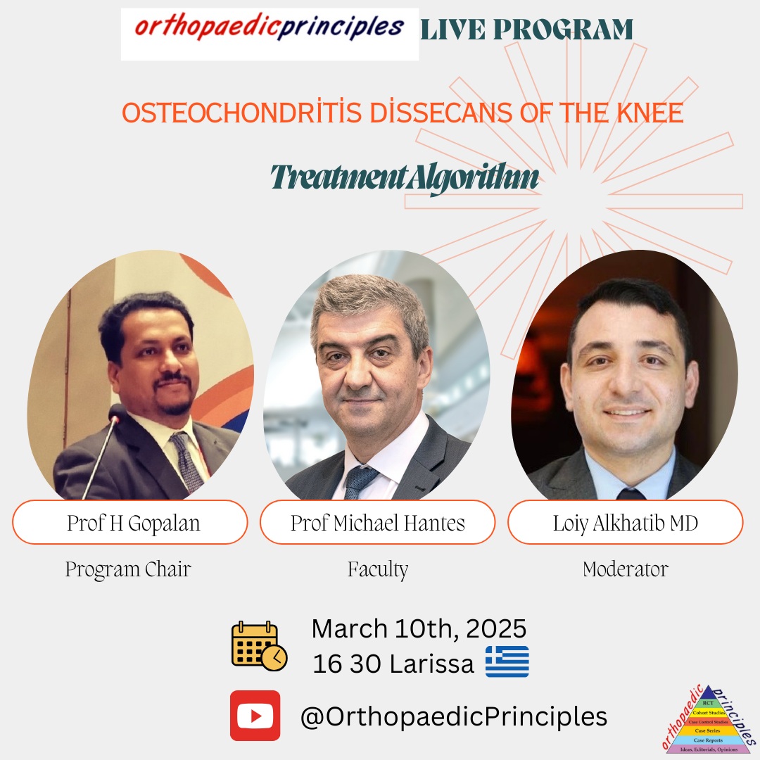
Courtesy: Prof Michael Hantes, ESSKA 1st Vice President
Lecture Topic: Osteochondritis Dissecans of the Knee
-
Aim: Define the treatment algorithm for this disease.
-
Overview:
-
Pediatric disease affecting the subchondral bone.
-
If it progresses, it may lead to instability and cartilage disruption, potentially causing premature osteoarthritis.
-
Can occur in elbow, knee, hip, but most commonly affects the knee.
-
Location of Lesions in the Knee
-
Most common site: Medial femoral condyle (75-80%), close to the notch.
-
Other locations:
-
Lateral femoral condyle (15%).
-
Patella (5-10%).
-
Tibia (<1%) (very rare).
-
Clinical Presentation & Diagnosis
-
Symptoms:
-
Knee pain after prolonged activity.
-
Crepitus, catching, or locking (if loose body is present).
-
Swelling in some cases.
-
No pathognomonic signs for diagnosis.
-
-
Physical Examination:
-
Limp, alignment, joint palpation, range of motion, joint effusion.
-
Wilson’s test (low sensitivity, not widely used).
-
Imaging & Classification
-
X-rays:
-
Can show osteochondral lesions or loose body fragments.
-
-
MRI (Highly Recommended):
-
Most informative tool (90% sensitivity, nearly 100% specificity).
-
Helps determine size, stability, and prognosis.
-
-
MRI-Based Hefti Classification:
-
Determines whether a lesion is stable or unstable.
-
Stable: Small signal changes, no fluid between fragment and bone.
-
Unstable: Fluid completely surrounding fragment, partially/completely detached fragment.
-
-
Arthroscopic Classifications:
-
Guhl Classification:
-
Stage 1: Softened cartilage, intact surface.
-
Stage 2: Early separation without detachment.
-
Stage 3: Partially detached lesion.
-
Stage 4: Loose body or cartilage defect.
-
-
ROCK Study Group Classification:
-
Stable lesions: “Cue ball” & “Shadow”.
-
Unstable lesions: “Wrinkle in the rug,” “Locked door,” “Trapdoor,” “Crater.”
-
-
Natural History & Prognosis
-
Lesions can heal or worsen spontaneously.
-
Prognostic Factors:
-
Younger age = better prognosis.
-
Smaller lesions = better healing potential.
-
Stable lesions heal better than unstable ones.
-
-
Treatment depends on:
-
Age, lesion size, symptoms, and patient expectations.
-
Non-Operative Treatment (Conservative Management)
-
Recommended for young patients (?13 years old) with stable, small lesions and mild symptoms.
-
Treatment approach:
-
Activity modification for 3-6 months.
-
Avoidance of high-impact sports.
-
X-ray/MRI follow-up every 3-6 months.
-
Immobilizers not recommended (healing is independent of bracing).
-
-
Success Rate:
-
Some cases heal completely in 6 months.
-
Others progress, requiring surgical intervention.
-
Surgical Treatment (When Required)
-
Indicated for:
-
Unstable lesions on MRI.
-
Mechanical symptoms (clicking, locking, swelling).
-
Failure of conservative management.
-
-
Goals:
-
Maintain joint congruity.
-
Fix unstable fragments.
-
Address the subchondral bone to promote healing.
-
Surgical Techniques
-
Drilling (Stimulation of Healing):
-
Antegrade (Transarticular):
-
Simple, direct access.
-
Violates cartilage, which may not be ideal.
-
-
Retrograde (Through Bone, Avoids Cartilage Violation):
-
More technically demanding.
-
Requires fluoroscopy guidance.
-
Avoids growth plate damage in young patients.
-
-
-
Fixation (For Partially Detached Lesions):
-
Metal Screws:
-
Effective but require removal later.
-
-
Bioabsorbable Pins/Screws:
-
Preferred choice (no need for removal).
-
High success rate (80-95%).
-
-
Example Procedure:
-
Retrograde drilling using an ACL guide.
-
Placement of 3-4 bioabsorbable pins for fixation.
-
-
-
Other Options for Large, Detached Lesions:
-
Mosaicplasty (Osteochondral Autograft Transplantation – OATS).
-
Bone grafting (if necessary).
-
Surgical Outcomes & Literature Review
-
Healing Rate:
-
90-95% success with retrograde drilling and fixation.
-
Average healing time: 3-4 months (can range from 6 weeks to 2 years).
-
-
Clinical Study Findings:
-
40 patients treated with bioabsorbable pins.
-
Healing observed in 90% of cases.
-
No major complications or need for secondary surgery.
-
Conclusion
-
Key Takeaways:
-
Early diagnosis and MRI evaluation are crucial.
-
Stable lesions can often heal with conservative management.
-
Unstable lesions require surgery, with drilling and fixation being highly successful.
-
Bioabsorbable fixation offers excellent outcomes without the need for screw removal.
-
-
Final Message: Proper patient selection and treatment algorithm lead to optimal outcomes in osteochondritis dissecans of the knee.
Osteochondritis Dissecans of the Knee – Surgical Techniques & Management Algorithm
Considerations for Surgical Techniques
-
Each technique has advantages and limitations.
-
Factors to consider:
-
Cost of the procedure.
-
One-step vs. two-step procedure.
-
Patient history and literature-based outcomes.
-
Alternative Surgical Options
Microfracture Technique
-
Previously suggested for these types of lesions.
-
Rarely used today due to poor long-term outcomes.
Fresh Osteochondral Graft
-
Potential option if available in certain countries.
-
Challenges:
-
Limited availability in most countries.
-
Lower success rate based on literature.
-
Risk of chondrocyte loss in long-term follow-ups.
-
High rate of reoperation in patients who undergo this method.
-
Mosaicplasty (Osteochondral Autograft Transfer – OATS)
-
Highly effective for smaller defects (<3-4 cm²).
-
Limitations:
-
Donor site morbidity can be a concern for larger defects.
-
Autologous Chondrocyte Implantation (ACI)
-
Considered one of the best options.
-
Limitations:
-
Expensive.
-
Two-stage procedure.
-
Not available in all countries.
-
Newly Developed Treatment Approach
Impaction Bone Grafting with Autologous Matrix-Induced Membrane
-
Developed as an alternative to overcome limitations of other techniques.
-
Procedure Steps:
-
Identify the lesion and debride the area.
-
Microfracture the bony bed to promote healing.
-
Harvest bone graft from the medial or lateral femoral condyle.
-
Impact the bone graft into the defect.
-
Place a hyaline-based membrane over the defect.
-
Stabilize the membrane with fibrin glue.
-
-
Key Advantages:
-
Overcomes fresh osteochondral allograft limitations.
-
Reduces concerns related to tissue availability, size matching, and cost.
-
No severe complications reported.
-
Surgical Procedure Breakdown
-
Pre-Operative Case Example:
-
27-year-old patient with unstable osteochondral lesions.
-
Two osteochondral fragments removed.
-
-
Arthroscopic Debridement:
-
Borders of the lesion are clearly defined.
-
Microfracture performed after ensuring a bleeding bone bed.
-
-
Bone Graft Harvesting:
-
Harvested from the medial or lateral femoral condyle.
-
Defect is prepared and filled with impacted bone graft.
-
-
Membrane Placement & Fixation:
-
Membrane cut to defect size using a template.
-
Fibrin glue applied to secure it in place.
-
Ensured that scaffold remains slightly lower than the cartilage surface.
-
-
Final Steps:
-
Joint movements checked before finalizing the procedure.
-
Ensured the membrane stays in position as fibrin glue reaction completes.
-
Clinical Outcomes & Literature Review
-
Study Data:
-
25 patients with a mean follow-up of 4 years.
-
Significant improvement in clinical scores pre-op vs. post-op.
-
-
MRI Findings:
-
Most cases showed healed subchondral bone and cartilage restoration.
-
MOAKS MRI scoring system used for assessment:
-
Higher MOAKS scores correlated with better clinical outcomes.
-
Despite moderate MOAKS scores, patients experienced significant clinical improvements.
-
-
-
Publication Reference:
-
Full details of the technique and study published in the “Chesta” journal.
-
Management Algorithm for Osteochondritis Dissecans
-
Treatment depends on:
-
Stage of the disease.
-
Patient age.
-
Activity level.
-
Available surgical options.
-
-
Recommended Approach:
-
Arthroscopic drilling and internal fixation for Stage 2 and 3 lesions.
-
Bone grafting with impaction and autologous matrix-induced chondrogenesis for larger defects.
-
-
Comparison with Other Techniques:
-
Mosaicplasty & Autologous Chondrocyte Implantation (ACI) are valid options, but cost and donor-site concerns must be considered.
-
The new impaction bone grafting technique is time- and cost-effective with promising results.
-
Upcoming Events & Professional Membership Information
-
Upcoming ESSKA Congress:
-
Dates: May 20-22, 2026.
-
Location: Prague, Czech Republic.
-
Looking forward to welcoming participants!
-
-
Becoming an ESSKA Member:
-
Open to surgeons worldwide, not just in Europe.
-
One-third of members are from outside Europe.
-
Visit the ESSKA website to apply.
-
-
European Certification Program:
-
Certifies surgeons in ACL, shoulder arthroscopy, patellofemoral instability, and other specialties.
-
Closing Remarks
-
Thank you to Professor Gopalan for the invitation.
-
Thank you to all participants for their attention!

Leave a Reply