OrthopaedicPrinciples.com

Integrating Principles and Evidence

Integrating Principles and Evidence

  • Home
  • Editorial Board
  • Our Books
    • Evidence Based Orthopaedic Principles
  • Courses
  • Exams
  • Reviews
  • Live Program
  • Contact

Anatomy of the Elbow

Courtesy: Prof Nabil Ebraheim, University of Toledo, Ohio, USA


Anatomy of the Elbow
Elbow is made of following bones
Humerus, Radius and Ulna

  • The lower end of the humerus have two epicondyles: lateral epicondyle and medial epicondyle
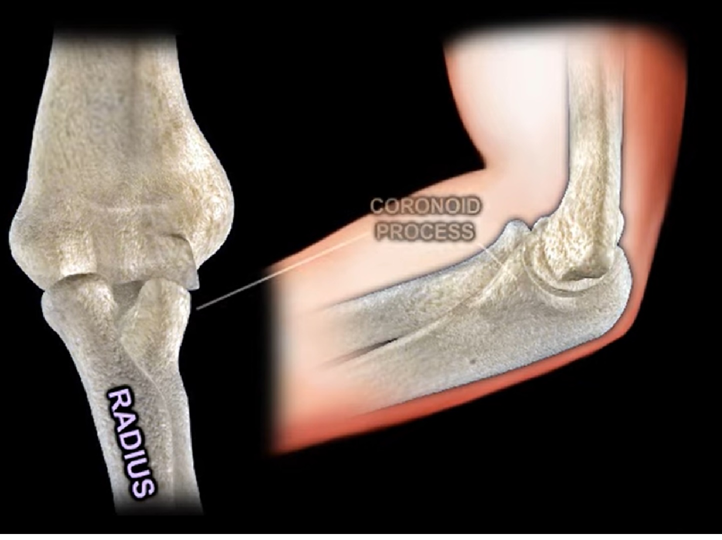
    and it also has the capitellum laterally and the trochlea medially
  • The proximal ulna is made of the olecranon process, the trochlear notch and the coronoid process.
  • The bursa lies on top of the olecranon process.
  • The radius is made of the radial head , neck and radial tuberosity .
  • The primary stabilizer of the elbow is the ulnohumeral joint .
  • The coronoid process is the anterior buttress of the olecranon it prevents posterior dislocation of the elbow.
  • fracture of the coronoid process more than 50 percent of the height will lead to elbow instability

Ligaments : Medial Elbow

Medial (ulnar) collateral ligament

MCL is composed of three bands

  • Anterior
  • Posterior
  • Transverse Bundles
  • The anterior band of MCL is the strongest primary stabilizer to valgus stress in 90 degree of flexion
  • Medial Collateral Ligament originates from the posterior medial epicondyle of the distal humerus and it inserts into the sublime tubercle of the medial coronoid process
  • Late cocking and early acceleration will give the highest valgus torque to the medial collateral ligament
  • Deficiency of this ligament is diagnosed with valgus on moving valgus stress test.
  • MRI: best study to diagnose complete tear of the MCL of the elbow
  • The posterior bundle of the MCL is tight in elbow flexion
  • To get more flexion of the stiff elbow, release the contracted posterior band of the medial collateral ligament

Muscles On The Medial Side Of The Elbow

Pronator teres muscle and the flexor muscle group of the forearm attached to the medial epicondyle
Flexor muscle :
• Biceps Major
• Flexor Carpi Radialis
• Palmaris longus
• Flexor Carpi Ulnaris
• Triceps Brachii

Medial Elbow Nerve: Ulnar nerve
• Location : Cubital Tunnel

Posterior Elbow Muscle

  • Triceps Major

Lateral Side Ligaments

  • Lateral Collateral Ligament
  • Lateral ulnar collateral ligament
  • Accessory lateral collateral ligament
  • Annular ligament


  • Lateral ulnar collateral ligament is the key anatomic structure which prevents posterolateral instability
  • The lateral ulnar collateral ligament acts like a sling for the radial head.
  • It is located in the posterolateral aspect of the radial head.
  • It arises from the lateral humeral epicondyle and it inserts into the crista supinatoris of the proximal ulna .
  • The posterolateral rotatory instability of the elbow occurs with the lateral ulnar collateral ligament deficiency.
  • It is diagnosed in a lateral pivot shift test
  • Impaction fracture of the anteromedial coronoid facet is usually associated with LCL injury
  • This will lead to posteromedial instability of the elbow
  •  Sometimes this fracture is not easily seen on x-rays and it can be missed
  • Fracture of the coronoid process can also be a part of the terrible elbow triad ( an elbow dislocation associated with a coronoid fracture and radial head fracture)

Lateral Side Muscles

  • extensor tendon muscles
  • Extensor carpi radialis longus
  • Extensor carpi radialis brevis
  • Extensor carpi ulnaris
  • Extensor digitorum

Anterior Elbow Anatomy

  • lateral to the biceps tendon is the radial nerve
  • medial to the biceps tendon in the brachial artery and median nerve
  • Biceps is inserted into the radial tuberosity and above the biceps lies the radial recurrent arteries
  • Branch from the musculocutaneous nerve can be commonly injured during distal biceps surgery

Post Views: 1,976

Related Posts

  • MRI Anatomy of the Elbow

    Courtesy: Dr Jean Jose MD Associate Chief Musculoskeletal Radiology Section Associate Professor of Clinical Radiology…

  • Biceps Brachii Anatomy

    Courtesy : Prof Nabile Ebraheim, University of Toledo, Ohio, USA

  • Acetabular fractures: Applied Anatomy

    Courtesy: Saqib Rehman MD Director of Orthopaedic Trauma Temple University Philadelphia Pennsylvania USA

Leave a Reply Cancel reply

Your email address will not be published. Required fields are marked *

Follow Us

instagram slideshare

Categories

  • -Applied Anatomy
  • -Approaches
  • -Basic Sciences
  • -Cartilage & Meniscus
  • -Classifications
  • -Examination
  • -Foot and Ankle
  • -Foot and Ankle Trauma
  • -FRCS(Tr and Orth) tutorials
  • -Gait
  • -Hand and Wrist
  • -Hand and Wrist Trauma
  • -Hand Infections
  • -Hip and Knee
  • -Hip Preservation
  • -Infections
  • -Joint Reconstruction
  • -Knee Arthroplasty
  • -Knee Preservation
  • -Metabolic Disorders
  • -Oncology
  • -OrthoBiologics
  • -OrthoPlastic
  • -Paediatric Orthopaedics
  • -Paediatric Trauma
  • -Patellofemoral Joint
  • -Pelvis
  • -Peripheral Nerves
  • -Principles
  • -Principles of Surgery
  • -Radiology
  • -Rheumatology
  • -Shoulder and Elbow
  • -Shoulder and Elbow Arthroplasty
  • -Spine Deformity
  • -Spine Oncology
  • -Spine Trauma
  • -Spine, Pelvis & Neurology
  • -Sports Ankle and Foot
  • -Sports Elbow
  • -Sports Knee
  • -Sports Medicine
  • -Sports Medicine Hip
  • -Sports Shoulder
  • -Sports Wrist
  • -Statistics
  • -Technical Tip
  • -Technology in Orth
  • -Trauma
  • -Trauma (Upper Limb)
  • -Trauma Life Support
  • -Trauma Reconstruction
  • Book Shelf
  • Book Shelf Medical
  • Careers
  • Case Studies and Free Papers
  • DNB Ortho
  • Evidence Based Orthopaedic Principles
  • Evidence Based Orthopaedics
  • Exam Corner
  • Fellowships
  • Guest Editor
  • Guest Reviews
  • Image Quiz
  • Instructional Course Lectures
  • Journal Club
  • MCQs
  • Meetings and Courses
  • Multimedia
  • News and Blog
  • Plaster Techniques
  • Podcasts
  • Public Health
  • Rehabilitation
  • Research
  • Shorts and Reels

Book Shelf

Kendall’s Muscle Testing and Function 6th Edition

Kendall’s Muscle Testing and Function 6th Edition

By admin Leave a Comment

Get Book Kendall’s Muscles: Testing and Function, with Posture and Pain, 6th Edition, transforms this landmark Physical Therapy classic to prepare you for unparalleled clinical success in today’s practice. Timeless coverage of manual muscle testing, evaluation, and treatment meets the latest evidence-based practices, engaging imagery, and dynamic digital resources to create a powerful resource you […]

Popular Posts

  • Bone Screws in Orthopaedic Surgery
  • Silverskold Test
  • Piriformis Syndrome
  • Blood Supply of Long Bone
  • Movements of the Thumb

Recent Comments

  • RAJATABHA BISWAS on NUH Fellowship in Singapore
  • Runj on ESSKA Congress 2026
  • OT Hand Therapist on Interosseous Muscles Of The Hand
  • Badreddine on Rockwood and Green Fractures in Adults and Children- 10th Edition
  • Prof Dr P.sridhar MS Ortho,D Ortho on Rockwood and Green Fractures in Adults and Children- 10th Edition

Exam Corner

FRCS Orth Exam- Knee Arthroplasty

Courtesy: Zaid al Rab, FOunder, OrthoPass

MCQ Exam for the FRCS Orth 1

Courtesy: Zaid al Rub, Founder, OrthoPass

Postgraduate Entrance Exam Set 3

Get explanatory answers from our book

Postgraduate Entrance Exam Set 2

Get explanatory answers from our book

Main Menu

  • Orthopaedic Principles
  • Editorial Board
  • Orthopaedic Principles-A Review

Recent Posts

  • Anterior Tibial Tendon Tears
  • Endoscopic Lumbar Microdiscectomy
  • RAMP Lesion of the Knee
  • Osteochondritis Dissecans of the Knee
  • Dual Mobility Cups in Total Hip Replacement

Links

  • Join Our Editorial Board
  • Journals
  • Weblinks
  • Submit Your Conference
  • Disclaimer
Copyright@orthopaedicprinciples.com. All right rerserved.