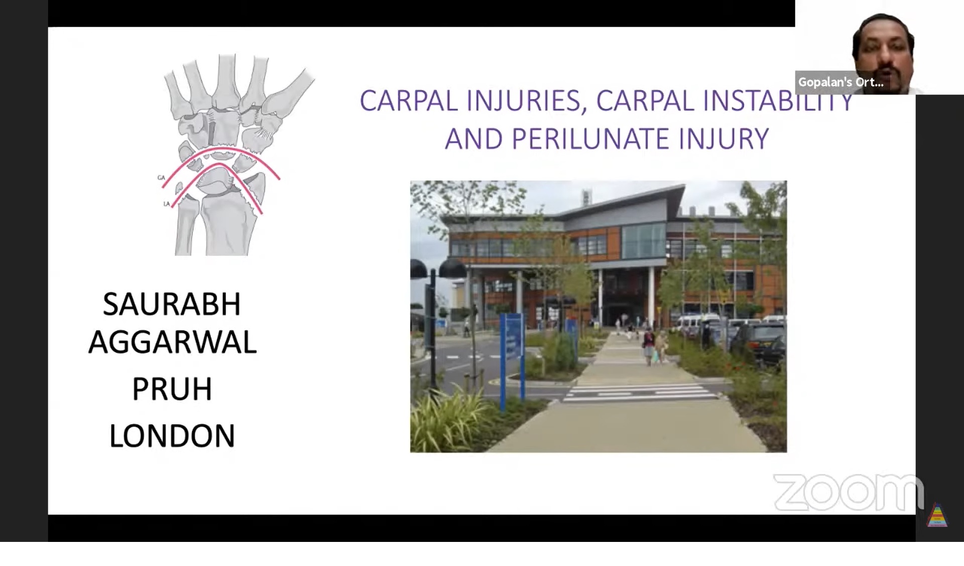
Courtesy: Saurabh Agarwal, Consultant, UK
PERILUNATE DISLOCATION
INTRODUCTION
• Perilunate injuries refer to disruptions in the ligamentous and bony connections of the lunate with adjacent carpal bones and the radius.
• These are severe wrist injuries that, if untreated, lead to long-term dysfunction and pain.
• Perilunar or perilunate dislocations account for the majority of wrist dislocations.
Anatomy & Gilula’s Lines
• Gilula’s Lines: Three smooth carpal arcs visible on a lateral X-ray to evaluate carpal alignment.
o Proximal convexity of scaphoid, lunate, and triquetrum.
o Distal convexity of the same bones.
o Proximal surfaces of capitate and hamate.
• Disruption indicates injury.
Mechanism of Injury
• High-energy trauma (e.g., falls, motor vehicle accidents).
• Force transmission through hyperextended wrist with varying intensity.
STAGES OF PERILUNATE INJURY (Mayfield Classification)
• Reverse pattern: Starts at Stage IV and progresses backward.
1. Stage 1: Scapholunate dissociation.
2. Stage 2: Lunocapitate dislocation.
3. Stage 3: Perilunate dislocation.
4. Stage 4: Lunate dislocation.
TYPES OF PERILUNATE INJURIES
Injury Patterns
• Common Patterns:
o Trans-scaphoid Perilunate Dislocation: Most frequently seen; includes scaphoid fracture and perilunate dislocation.
• Variability:
o Partial or complete lunate dislocation.
o Independent displacement of fractured carpal bones.
Grades of Lunate Dislocation
1. Grade 1: Normal alignment.
2. Grade 2: Palmar rotation 90° but attached to the radius.
4. Grade 4: Complete enucleation without radius attachment.
Clinical Presentation
• Symptoms:
o Pain, swelling, and deformity (dinner-fork appearance).
o Radial deviation of the wrist.
• Physical Signs:
o Palpable lunate prominence volarly.
o Tenderness over the wrist and reduced motion.
• Complications:
o Median nerve compression from volar lunate dislocation.
Diagnostic Imaging
• Radiographic Features:
o Loss of normal Gilula’s arcs.
o “Spilled teapot sign” in lateral view: Volar tilting of lunate.
o Triangular lunate shape in AP view.
o Capitate misalignment with lunate fossa.
o Associated fractures (scaphoid, triquetrum).
• Advanced Imaging:
o CT scan: For fracture detail.
o MRI: To evaluate ligament integrity.
Treatment
Closed Reduction
• Principle: Restore alignment without surgery.
• Steps:
1. Axial traction.
2. Flexion of the wrist.
3. Restoration of carpal alignment.
• Success Criteria:
o Restored Gilula’s arcs.
o Symmetric scapholunate gap.
o Normal intercarpal relationships.
• Immobilization: Cast for 8–12 weeks.
Pinning
• Percutaneous pinning to stabilize joints during healing.
Surgical Management
• Preferred Approach: Open Reduction + Internal Fixation (ORIF).
Complications and Outcomes
• Complications:
o Chronic instability.
o Post-traumatic arthritis.
o Median nerve damage.
• Long-Term Outcomes: Dependent on early diagnosis and reduction quality.
Summary
• Early recognition and staged understanding of perilunate injuries are critical.
• Treatment depends on severity:
o Closed reduction for minor injuries.
o ORIF for severe cases.
• Post-reduction care is essential for functional recovery.

Leave a Reply