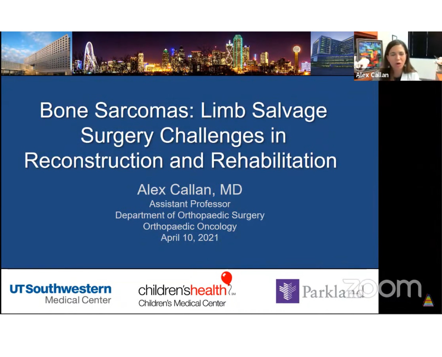
Courtesy: Alex Callan, MD, Assistant Professor, Orthopaedic Oncologist, UT SouthWestern Medical Centre, Dallas, Texas
Bone Sarcomas: Limb Salvage Surgery Challenges in Reconstruction and Rehabilitation
Objectives:
1. Describe bone sarcoma etiology and epidemiology
2. Understand treatment algorithms for bone Sarcomas in kids
3. List options for local control surgery (the 4 As!) : Amputation – Allograft – Arthroplasty – APC)
4. Understand rehabilitation challenges in limb salvage
SARCOMAS
- Origin: Greek “sarx” = flesh
- Sarcoma is a Cancer from a mesenchymal Cell line
- These are malignant tumors arising From bone, cartilage, muscle, Fat, nerve or vessels
MALIGNANT BONE TUMORS IN KIDS
Approximately 1200 newly diagnosed bone Sarcomas annually
-Osteosarcoma (most common)
-Ewing Sarcoma
Location
OSTEOSARCOMA
Common location is METAPHYSIS mainly in
-Distal Femur (40%)
– Proximal Tibia (20%)
-Proximal Humerus (10%)
EWING SARCOMA
Common location is diaphysis
Pelvis & scapula are favoured sites
OSTEOSARCOMA
- Metaphyseal or metadiaphyseal
- Locations : Distal femur, proximal tibia, proximal femur and humerus
- Codman’s triangle where new bone forms in response to periosteal elevation
- Sunburst` appearance when the periosteum
- Does not have enough time to lay down a new layer and instead the Sharpey’s fibres stretch Perpendicular to the periosteum
TYPES
Conventional Intramedullary Osteosarcoma
Non-conventional
— Parosteal Osteosarcoma
– Periosteal Osteosarcoma
– Telangectatic Osteosarcoma
– High Grade Surface OS or Low Grade Intramedullary
Small Cell Osteosarcoma
Secondary Osteosarcoma
Conventional Osteosarcoma
– Osteoblastic osteosarcoma
– Chondroblastic osteosarcoma
– Fibroblastic osteosarcoma
Conventional Osteosarcoma
– Overall, if >90% necrosis
75% 5-year survival
70% 10-year survival
If Necrosis is less than 90%, worse survival
EWINGS SARCOMA
- Presenting complaints are Pain, fevers, swelling
- Age: 10-30s
- Radiographs: Sunburst appearance, Onion Skin appearance, Codman triangle
- Common location is diaphyseal or flat bone
- Diagnosis : Biopsy -> Small round blue Cells– FISH -> Translocation (t 11;22) ESW:FLI1
- Staging Work-up:
Xrays entire Bone, MRI entire Bone, CXR
Chest CT, PET Scan, Bone Marrow Biopsy - PATHOLOGY
Sheets of blue, round cells
Large nuclei (blue)
Scant cytoplasm (primitive)
Glycogen +
CD99 +
No extracellular matrix
Treatment:
Diagnosis and Staging
– Biopsy
– MRI extremity, CT Chest, Bone Scan or PET Scan
Chemotherapy (Neoadjuvant) for 10-12 weeks
– Non Weight bearing Extremity to prevent fracture
Surgery (Radical Resection & Reconstruction)
Chemotherapy (Adjuvant)
Surveillance
Secondary goals
- Provide the best reconstruction to restore Function to the limb
- This is complicated with tumors located in Close proximity to joints, muscles and Neurovascular structures.
- To make matters worse many patients are still growing kids.
Limb Salvage Surgery is performed if:
- Adequate margin for resection of tumor can be obtained with low risk ( 8 cm Extensive muscle or soft-tissue involvement
- Poor response to preoperative chemotherapy
The various modalities for limb reconstruction used are:
Arthrodesis
Mobile joint reconstruction
– Autoclaved tumor bone
– Allograft bone including osteoarticular allograft
– Bone transfer (ulna and fibula to reconstruct radius and tibia respectively), Huntington’s procedure
– Endoprosthetic reconstruction
–Allograft end prosthetic reconstruction
– Custom-made prosthesis
– Rotationplasty
Amputation
- Above Knee Amputation (AKA)
- Hip Disarticulation
- External Hemipelvectomy
Rotationplasty
Limb-Salvage Treatment versus Amputation for Osteosarcoma Of the Distal End of the Femur:
Challenges with MEGA joint replacement
- Surgically removal all soft tissue Attachments (muscle, tendon, ligaments)
- Attempt to re-attach to metal implant or Cadaver bone
- Quad weakness, extensor lag, hip girdle Dysfunction, cuff atrophy
- Nerve palsies
- Balance between Healing and Motion
Complex, Personalized Rehabilitation Based on Reconstruction Technique
May Need Prolonged Joint Immobilization to allow healing
Keep leg in a functional position
– Knee straight (avoid flexion contracture)
– Foot Plantigrade (avoid equinus contracture)
May need prolonged Non Weight Bearing status to allow healing to implant or allograft
Distal Femur Endoprosthesis:
- Cemented Implants allow for Immediate Weight Bearing
- Usually – Weight bearing as tolerated, no brace, no Restrictions
- If massive quadriceps resection, may use Hinged knee brace for ambulation Until quad strength returns
Proximal Tibia Endoprosthesis
- No Knee ROM! (KneeBrace locked in extension X 6 wks
- 3D walker boot
- Progressive ROM x 6 wks
Proximal Femur Endoprosthesis
Used in Anterior/Posterior Hip dislocation
Precautions :
No flexion over 80
No adduction
+/- Hip abduction Brace
+/- Knee lmmobilizer
Proximal Humerus/Total Humerus Endoprosthesis
Non weight bearing
Use Sling for 6-12 wks
No active motion to shoulder
Repair rotator cuff to metal
Limb Salvage Surgery : Distal femur COMPRESS Endoprosthesis
Need for Bony Ingrowth onto Implant
Work on Quad Strength and Knee ROM
Solutions for the Growing Child :
- Preserve Physis if possible
- Intercallary allograft
- Shut down contralateral physis
- Osteoarticular allograft
- Growing endoprosthesis
– Minimally invasive expandable - Non-invasive, magnetic expandable
– Girls reach skeletal maturity 12-14yr
– Boys reach skeletal maturity 14-16yr
Consider involved physis
Physeal Growth: Proximal Femur 3mm/yr, Distal femur 9mm/yr, Proximal tibia 6mm/yr, Distal tibia 5mm/yr
Reconstruction options
- Amputation (AKA or knee disarticulation)
- Osteoarticular allograft
- Arthroplasty: Proximal Tibial Endoprosthesis
Standard
– Minimally Invasive Expandable
– Magnetic, non-invasive expandable APC – allograft prosthetic complex
Proximal tibia allograft, hinged TKA
Management:
- Knee Immobilizer -> Cast
- Crutches or walker
- Non-Weight bearing
- Pain Control
Staging and Diagnosis
- X-ray entire Bone
- MRI Tibia with and without contrast
- Chest Xray
- CT Chest with contrast
- Whole Body Bone Scan
- Biopsy and cast placement
Summary:
- Bone Sarcomas (Osteosarcoma and Ewing Sarcoma are rare! 1200 new cases each year)
- Bone sarcoma treatment: Chemo – Surgery-Chemo
- List options for local control surgery (the 4 As!
Amputation – Allograft – Arthroplasty – APC) - Rehabilitation and recovery are challenging in limb salvage

Leave a Reply