OrthopaedicPrinciples.com

Integrating Principles and Evidence

Integrating Principles and Evidence

  • Home
  • Editorial Board
  • Our Books
    • Evidence Based Orthopaedic Principles
  • Courses
  • Exams
  • Reviews
  • Live Program
  • Contact

Kienbock’s disease

Courtesy: Prof Abhijeet Wahegaonkar, Sancehti Hospital, Pune, India

Introduction
• Robert Kienbock- radiologist. Described xray changes associated with lunatomalacia.
• an isolated disorder of the carpal lunate arising out of vascular compromise
• multifactorial pathogenesis- influenced by genetic, anatomic, mechanical and metabolic factors.

Historical perspectives

  • first described by Peste in 1843 who noted a collapsed lunate in cadaver dissection
  • Robert Kienbock- radiologist. Described Xray changes associated with
    Kienböck’s disease

Etiology

  • etiology is still undefined so various factors have been proposed
  • aberrant blood supply to lunate: problems with arteries[single dominant nutrient arterial supply for lunate (20%patients)
  • poorly organized intraosseous circulation —“i”pattern of anastomosis
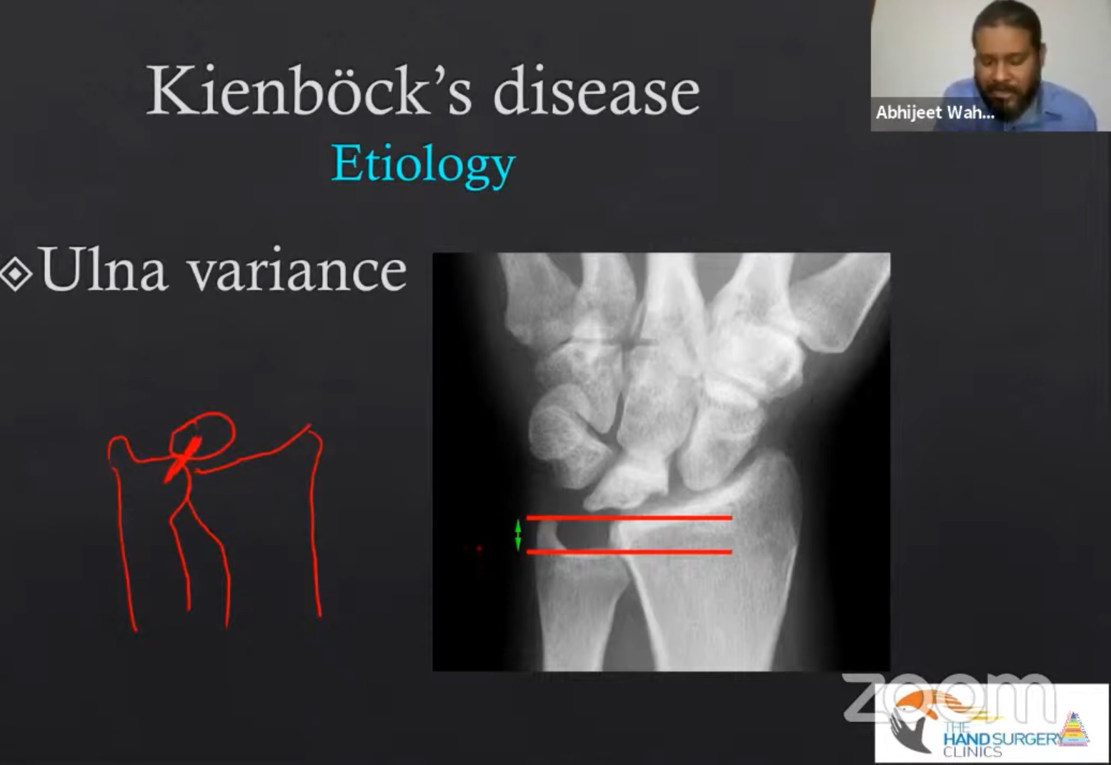
  • ulnar variance (radioulnar index) = distance between distal point of ulna on radial side and the distal point of radius on ulnar side. Normal is ±2, -2 means ulna is 2 mm shorter than radius.
  • lunate geometry
  • mechanical injury

Diagnosis:

  • symptoms can vary depending upon the stage at initial presentation.
  • pain localised to the radiolunate facet pain is classically insidious in onset
  • pain and stiffness in the dominant wrist
  • decreased grip strength
    hyperextension injury

Radiographs

X-ray imaging during stage 1 of the disease will show an apparently normal lunate bone, later sclerosis, fracture, fragmentation and collapse are seen.

Bone scan
o nonspecific
o does not rule out early AVN

Trispiral tomography
more accurate staging than x-ray alone
• does not rule out early AVN

CT scan
• provides useful detail of fracture fragments

Mri
• sensitive, specific

  • magnetic resonance imaging can help to assess the blood supply to the lunate.
  • The gadolinium-infused mri has been used to find the vascular patterns
    abnormal signal in 50% of lunate

Staging

  • Lichtman modification of Stähl’s classification
    • stage 1-4
    • modified by Amadio
  • stage 1- no visible changes on radiograph; changes seen on mri.
  • stage 2- sclerosis of lunate
  • stage 3a- fragmentation of lunate
  • stage 3b- fixed rotation of scaphoid
  • stage 4- degeneration of adjacent intercarpal joints

Treatment

  • based on the stage at presentation
  • unload the lunate
  • revascularise the lunate
  • treat carpal instability and collapse with salvage procedure

Stage 1
• conservative treatment with 3 months immobilisation is typically recommended for stage 1 disease

  • the patient should continue to be monitored and if symptoms or radiographs progress consider surgical management.

Stage II or III with negative ulnar variance

  • goal in this stage is generally centered towards unloading of lunate is an attempt to reduce
    intracarpal stress and allow revascularization.

Joint levelling procedures-
• radial shortening osteotomies
• ulnar lengthening procedures

Radial osteotomy is preferred over ulnar due to less complication

Stage II and 3a ulnar neutral or positive variance

  • revascularisation
  • osteotomies
  • core decompression

Stage 3b
o goal in this stage
o stabilization of carpus
o prevent further collapse

Decrease the load across radiolunate joint

  • proximal row carpectomy
    • scaphotrapeziotrapezoid arthrodesis
    • scaphocapitate arthrodesis
    • grafting ,arthroplasty and interposition

Stage iv
• salvage procedures performed.
• wrist arthrodesis
• wrist arthroplasty
• wrist denervation.

Summary

  • Kienbock disease is defined by AVN of lunate, with a predictable pattern of lunate collapse .carpal changes, and degeneration resulting from an apparent combination of vascular, anatomical and traumatic insults


Post Views: 4,476

Related Posts

  • Kienbock's disease

    KIENBOCK DISEASE • It is the avascular necrosis of the lunate. Lunate is one of…

  • Dupuytren's Disease Simplified

    Courtesy: Charles Eaton MD, Dupuytrens Research Group Multimedia presentation on Dupuytren's Disease: History, Aetilogy, Pathogenesis,…

  • Needle Aponeurotomy for Dupuytren's Disease

    Courtesy: Chris Bainbridge, Pulvertaft Hand Centre, UK

Leave a Reply Cancel reply

Your email address will not be published. Required fields are marked *

Follow Us

instagram slideshare

Categories

  • -Applied Anatomy
  • -Approaches
  • -Basic Sciences
  • -Cartilage & Meniscus
  • -Classifications
  • -Examination
  • -Foot and Ankle
  • -Foot and Ankle Trauma
  • -FRCS(Tr and Orth) tutorials
  • -Gait
  • -Hand and Wrist
  • -Hand and Wrist Trauma
  • -Hand Infections
  • -Hip and Knee
  • -Hip Preservation
  • -Infections
  • -Joint Reconstruction
  • -Knee Arthroplasty
  • -Knee Preservation
  • -Metabolic Disorders
  • -Oncology
  • -OrthoBiologics
  • -OrthoPlastic
  • -Paediatric Orthopaedics
  • -Paediatric Trauma
  • -Patellofemoral Joint
  • -Pelvis
  • -Peripheral Nerves
  • -Principles
  • -Principles of Surgery
  • -Radiology
  • -Rheumatology
  • -Shoulder and Elbow
  • -Shoulder and Elbow Arthroplasty
  • -Spine Deformity
  • -Spine Oncology
  • -Spine Trauma
  • -Spine, Pelvis & Neurology
  • -Sports Ankle and Foot
  • -Sports Elbow
  • -Sports Knee
  • -Sports Medicine
  • -Sports Medicine Hip
  • -Sports Shoulder
  • -Sports Wrist
  • -Statistics
  • -Technical Tip
  • -Technology in Orth
  • -Trauma
  • -Trauma (Upper Limb)
  • -Trauma Life Support
  • -Trauma Reconstruction
  • Book Shelf
  • Book Shelf Medical
  • Careers
  • Case Studies and Free Papers
  • DNB Ortho
  • Evidence Based Orthopaedic Principles
  • Evidence Based Orthopaedics
  • Exam Corner
  • Fellowships
  • Guest Editor
  • Guest Reviews
  • Image Quiz
  • Instructional Course Lectures
  • Journal Club
  • MCQs
  • Meetings and Courses
  • Multimedia
  • News and Blog
  • Plaster Techniques
  • Podcasts
  • Public Health
  • Rehabilitation
  • Research
  • Shorts and Reels

Book Shelf

Kendall’s Muscle Testing and Function 6th Edition

Kendall’s Muscle Testing and Function 6th Edition

By admin Leave a Comment

Get Book Kendall’s Muscles: Testing and Function, with Posture and Pain, 6th Edition, transforms this landmark Physical Therapy classic to prepare you for unparalleled clinical success in today’s practice. Timeless coverage of manual muscle testing, evaluation, and treatment meets the latest evidence-based practices, engaging imagery, and dynamic digital resources to create a powerful resource you […]

Popular Posts

  • Bone Screws in Orthopaedic Surgery
  • Silverskold Test
  • Piriformis Syndrome
  • Blood Supply of Long Bone
  • Movements of the Thumb

Recent Comments

  • RAJATABHA BISWAS on NUH Fellowship in Singapore
  • Runj on ESSKA Congress 2026
  • OT Hand Therapist on Interosseous Muscles Of The Hand
  • Badreddine on Rockwood and Green Fractures in Adults and Children- 10th Edition
  • Prof Dr P.sridhar MS Ortho,D Ortho on Rockwood and Green Fractures in Adults and Children- 10th Edition

Exam Corner

FRCS Orth Exam- Knee Arthroplasty

Courtesy: Zaid al Rab, FOunder, OrthoPass

MCQ Exam for the FRCS Orth 1

Courtesy: Zaid al Rub, Founder, OrthoPass

Postgraduate Entrance Exam Set 3

Get explanatory answers from our book

Postgraduate Entrance Exam Set 2

Get explanatory answers from our book

Main Menu

  • Orthopaedic Principles
  • Editorial Board
  • Orthopaedic Principles-A Review

Recent Posts

  • Anterior Tibial Tendon Tears
  • Endoscopic Lumbar Microdiscectomy
  • RAMP Lesion of the Knee
  • Osteochondritis Dissecans of the Knee
  • Dual Mobility Cups in Total Hip Replacement

Links

  • Join Our Editorial Board
  • Journals
  • Weblinks
  • Submit Your Conference
  • Disclaimer
Copyright@orthopaedicprinciples.com. All right rerserved.