OrthopaedicPrinciples.com

Integrating Principles and Evidence

Integrating Principles and Evidence

  • Home
  • Editorial Board
  • Our Books
    • Evidence Based Orthopaedic Principles
  • Courses
  • Exams
  • Reviews
  • Live Program
  • Contact

Sciatic Nerve Course, Branches to Lower Leg and Foot

Courtesy: Prof Nabil Ebraheim, University of Toledo, Ohio, USA

Sciatic nerve course and branches to the leg and foot
• The sciatic nerve ends just above the popliteal fossa , by dividing it in to :
1. Common peroneal nerve
2. Tibial nerve
• Tibial nerve supplies the posterior muscles of the leg:

1. Medial and lateral head of the gastrocnemius
2. Soleus
3. Tibialis posterior
4. Flexor digitorum longus
5. Flexor hallucis longus
• The common peroneal nerve is a branch of the sciatic nerve that travels posterior to the tendon of biceps femoris and it passes across the neck of fibula.

It pierces the peroneus longus and then divides in to
1. Superficial peroneal nerve
2. Deep peroneal nerve

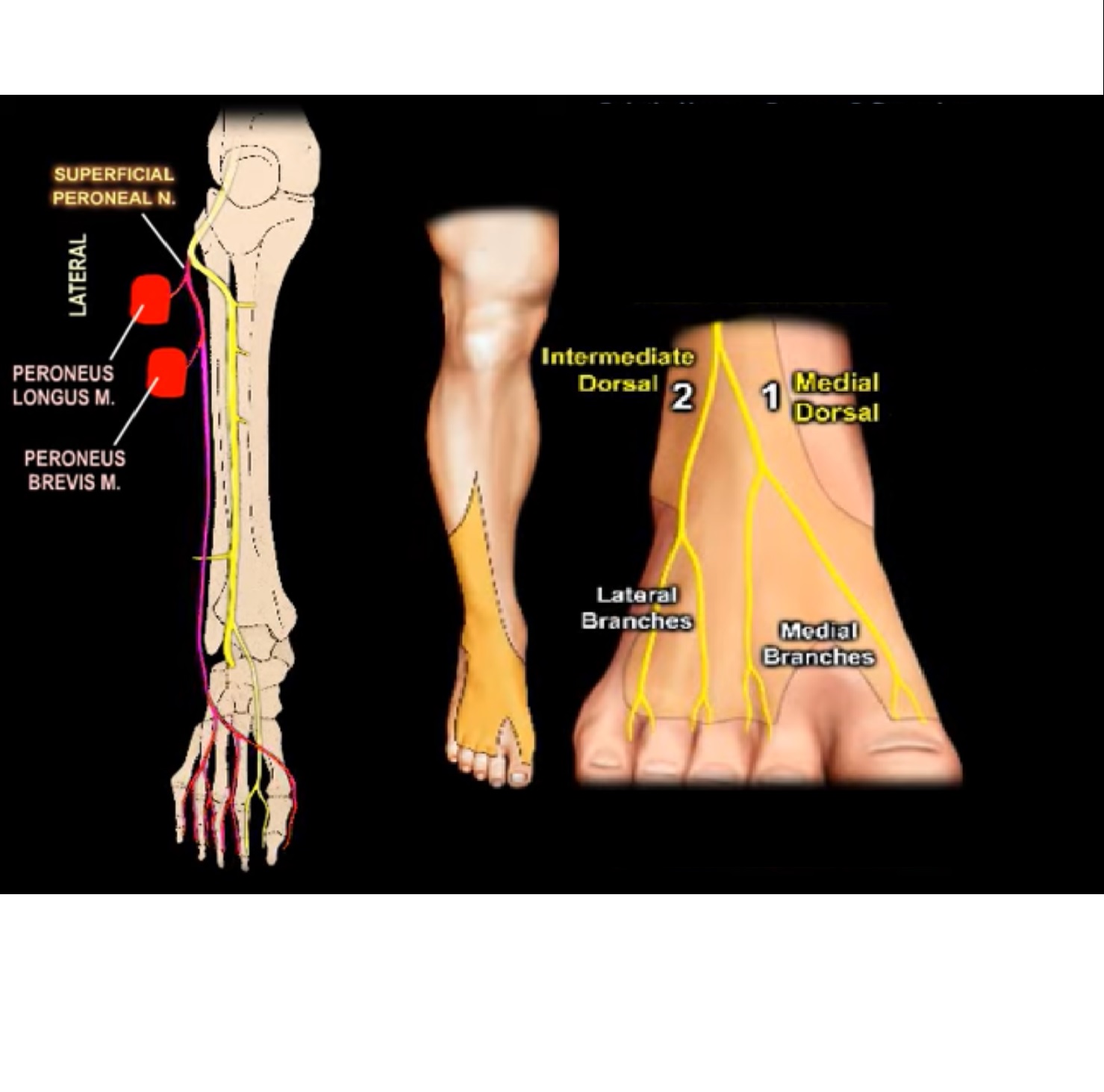
Superficial peroneal nerve:
• Supplies muscles of lateral aspect of leg– peroneus longus and peroneus brevis.
• Superficial peroneal nerve is called superficial or sub cutaneous because some part of the nerve go through the muscles and some parts are cutaneous
• In the proximal part of the leg the superficial peroneal nerve goes through the substance of the peroneus longus muscle .
• In the middle third of the leg the superficial peroneal nerve is located between the peroneus longus and peroneus brevis muscle.
• It is seen in the groove between the peroneus brevis and extensor digitorum longus muscle.
• In the distal 3rd of the leg the superficial peroneal nerve then pierces the fascia to become superficial .
• In the ankle the superficial peroneal nerve divides in to intermediate , dorsal cutaneous and medial dorsal cutaneous branches.
• Superficial peroneal nerve has a variable course and it becomes superficial to the peroneus longus muscle in the distal third of the leg.
• Superficial peroneal nerve divides in to sensory branches that supply the skin of the dorsum of foot.
• Function: eversion of foot
• Injured during fasciotomy of the lateral compartment of the leg

Deep peroneal nerve :
• Supplies the muscles of anterior compartment of the leg—tibialis anterior, extensor hallucis longus, extensor digitorum longus, peroneus tertius ,extensor digitorum brevis
• It gives a sensory branch to first web space, b/w 1st and 2nd toes – helps in diagnosing compartment syndrome
• Function: dorsiflexion of ankle and the toes
• Causes foot drop

Tibial nerve
• Passes in to foot , running posterior to the medial malleolus and beneath the flexor retinaculum
• It divides in to:
1. Medial and lateral plantar branches
2. Medial calcaneal branch
• Tibial nerve gives innervation to all muscles of foot except the extensor digitorum brevis
• Medial plantar nerve gives branches to
1. Abductor hallucis
2. Flexor digitorum brevis
3. Flexor hallucis brevis
4. First lumbricals
5. Digital nerves to medial 3 ½ toes
• Therefore medial plantar nerve innervation is similar to innervation of median nerve in the hand
• Lateral plantar nerves gives digital nerves to
1. Lateral 1 ½ toes
2. Abductor digiti minimi
3. Quadratus plantae
4. Adductor hallucis
• Similar to ulnar nerve in the hand, it gives branches to all interossei and from the 2nd to the 4th lumbricals
• It is the nerve which gives innervation to most intrinsic muscles of foot

• Baxters nerve: 1st branch of lateral plantar nerve: important in d/d of plantar fascitis


Post Views: 8,706

Related Posts

  • Nerves of the Lower Leg

    Courtesy: Prof Nabile Ebraheim, Professor of Orthopaedics and Traumatology, University of Toledo, Ohio, United States…

  • Superficial Peroneal Nerve Release in the Lower Leg

    Superficial Peroneal Nerve Release in the Lower Leg - Standard Authors: Mackinnon SE1, Yee A1…

  • Sciatic Nerve Injury after THR

    Courtesy: Dominic Power MD, Peripheral Nerve Surgeon, Birmingham, UK

Leave a Reply Cancel reply

Your email address will not be published. Required fields are marked *

Follow Us

instagram slideshare

Categories

  • -Applied Anatomy
  • -Approaches
  • -Basic Sciences
  • -Cartilage & Meniscus
  • -Classifications
  • -Examination
  • -Foot and Ankle
  • -Foot and Ankle Trauma
  • -FRCS(Tr and Orth) tutorials
  • -Gait
  • -Hand and Wrist
  • -Hand and Wrist Trauma
  • -Hand Infections
  • -Hip and Knee
  • -Hip Preservation
  • -Infections
  • -Joint Reconstruction
  • -Knee Arthroplasty
  • -Knee Preservation
  • -Metabolic Disorders
  • -Oncology
  • -OrthoBiologics
  • -OrthoPlastic
  • -Paediatric Orthopaedics
  • -Paediatric Trauma
  • -Patellofemoral Joint
  • -Pelvis
  • -Peripheral Nerves
  • -Principles
  • -Principles of Surgery
  • -Radiology
  • -Rheumatology
  • -Shoulder and Elbow
  • -Shoulder and Elbow Arthroplasty
  • -Spine Deformity
  • -Spine Oncology
  • -Spine Trauma
  • -Spine, Pelvis & Neurology
  • -Sports Ankle and Foot
  • -Sports Elbow
  • -Sports Knee
  • -Sports Medicine
  • -Sports Medicine Hip
  • -Sports Shoulder
  • -Sports Wrist
  • -Statistics
  • -Technical Tip
  • -Technology in Orth
  • -Trauma
  • -Trauma (Upper Limb)
  • -Trauma Life Support
  • -Trauma Reconstruction
  • Book Shelf
  • Book Shelf Medical
  • Careers
  • Case Studies and Free Papers
  • DNB Ortho
  • Evidence Based Orthopaedic Principles
  • Evidence Based Orthopaedics
  • Exam Corner
  • Fellowships
  • Guest Editor
  • Guest Reviews
  • Image Quiz
  • Instructional Course Lectures
  • Journal Club
  • MCQs
  • Meetings and Courses
  • Multimedia
  • News and Blog
  • Plaster Techniques
  • Podcasts
  • Public Health
  • Rehabilitation
  • Research
  • Shorts and Reels

Book Shelf

Kendall’s Muscle Testing and Function 6th Edition

Kendall’s Muscle Testing and Function 6th Edition

By admin Leave a Comment

Get Book Kendall’s Muscles: Testing and Function, with Posture and Pain, 6th Edition, transforms this landmark Physical Therapy classic to prepare you for unparalleled clinical success in today’s practice. Timeless coverage of manual muscle testing, evaluation, and treatment meets the latest evidence-based practices, engaging imagery, and dynamic digital resources to create a powerful resource you […]

Popular Posts

  • Bone Screws in Orthopaedic Surgery
  • Silverskold Test
  • Piriformis Syndrome
  • Blood Supply of Long Bone
  • Movements of the Thumb

Recent Comments

  • RAJATABHA BISWAS on NUH Fellowship in Singapore
  • Runj on ESSKA Congress 2026
  • OT Hand Therapist on Interosseous Muscles Of The Hand
  • Badreddine on Rockwood and Green Fractures in Adults and Children- 10th Edition
  • Prof Dr P.sridhar MS Ortho,D Ortho on Rockwood and Green Fractures in Adults and Children- 10th Edition

Exam Corner

FRCS Orth Exam- Knee Arthroplasty

Courtesy: Zaid al Rab, FOunder, OrthoPass

MCQ Exam for the FRCS Orth 1

Courtesy: Zaid al Rub, Founder, OrthoPass

Postgraduate Entrance Exam Set 3

Get explanatory answers from our book

Postgraduate Entrance Exam Set 2

Get explanatory answers from our book

Main Menu

  • Orthopaedic Principles
  • Editorial Board
  • Orthopaedic Principles-A Review

Recent Posts

  • Anterior Tibial Tendon Tears
  • Endoscopic Lumbar Microdiscectomy
  • RAMP Lesion of the Knee
  • Osteochondritis Dissecans of the Knee
  • Dual Mobility Cups in Total Hip Replacement

Links

  • Join Our Editorial Board
  • Journals
  • Weblinks
  • Submit Your Conference
  • Disclaimer
Copyright@orthopaedicprinciples.com. All right rerserved.