Courtesy: Prof Nabil Ebraheim, University of Toledo, Ohio, USA
COMMON CONDITIONS CAUSING LOW BACK PAIN
RISK FACTORS:
- Job dissatisfaction
- Twisting
- Excessive, continuous vibration at job
- Heavy weight lifting
- Stress on back / Stress and depression
- Cigarette smoking – As it affects the vascularity & nutrition of vertebral discs
RELATION BETWEEN INTRADISCAL PRESSURE (IDP) AND DIFFERENT BODY POSITIONS:
- Supine position gives the lowest Intradiscal Pressure- IDP (25 kg), whereas a person in sitting position leaning forward & holding weight gives the highest IDP (275 kg).
- Presence of multiple Waddell signs (like over reaction, exaggeration, tenderness to light touch) on examination of patient indicates a non organic symptom etiology.
HISTORY AND EXAMINATION OF PATIENT:
1) If the pain is worse with flexion (like with sitting), it indicates Disc prolapse.
2) If the pain radiates to leg, it implies Disc herniation.
3) If the pain is more in back, then it is attributed to discogenic back pain.
4) If the pain worsens with extension and is more of back pain, it indicates Spondylosis/ Spondylolisthesis.
5) If the pain worsen with extension & is more of leg pain than back pain implies Spinal stenosis (Narrowing of spinal canal or foramen).
DIAGNOSTIC CRITERIA OF HERNIATED DISC:
1) SLR test
2) Disc herniation on imaging study
3) Neurological findings
If all these are positive, there is 95% chance of herniated disc. If only SLR test and imaging study are positive, there is 85% chance of herniated disc. There is only 66% chance of herniated disc if only SLR test is positive.
STRAIGHT LEG RAISING TEST:
- Patient is kept in supine position with knee extended as he/she flexes the hip. It stretches the Sciatic nerve.
- At 35 – 70° flexion of hip, irritation of nerve root (L5-S1 nerve root) is mainly due to herniated disc..
- A positive contralateral SLR test is very specific for lumbar disc herniation.
IMAGING STUDIES:
It includes the following,
- X rays
- MRI: It is indicated in conditions like:
a. Pain for > 4 weeks which is not relieving in nature.
b. Suspect Cauda equina syndrome/ infection/ tumor/ trauma.
c. Ankylosing spondylitis/ DISH even with minor trauma. - MRI with gadolinium differentiates recurrent disc herniation from fibrosis, as the latter enhances with gadolinium whereas recurrent disc herniation does not.
MOST COMMON CAUSES OF LOW BACK PAIN:
1) Intervertebral disc disorders: Herniated disc, Internal disc disruption, Cauda equina syndrome, Degenerative disc disease
2) Lumbar stenosis
3) Spondylolisthesis
4) Degenerative scoliosis
5) Failed back syndrome
6) Sacroiliac joint dysfunction
HERNIATED DISC:
It mostly affect the L4-L5 and L5-S1 levels. It is characterized by back pain and leg pain which is radicular in type. Mostly occurs in the posterolateral aspect, affecting the traversing (descending/lower nerve root) nerve root, ie; L5 nerve root for L4-L5 disc prolapse.
In cases of far lateral disc herniation/ foraminal disc herniation, it affects the exiting nerve root (upper nerve root) in 5-10% cases.
SLR test is positive and is called the tension sign. Types of disc herniation vary from protrusion/ bulge to disc herniation to sequestration.
MANIFESTATIONS OF L4, L5, S1 NERVE ROOT IRRITATIONS:
L4: MOTOR- Knee extension (L2,L3,L4)
L4 ankle dorsiflexion (Tibialis anterior)
SENSORY- Medial side of leg down to medial side of foot
REFLEXES- Patellar reflex
L5: MOTOR- Hip abduction (Gluteus medius)
Extension of big toe
SENSORY- Dorsum of foot and leg
S1: MOTOR- Hip extension (Gluteus maximus)
Foot eversion (Peroneus longus and Peroneus brevis)
Ankle plantar flexion (Gastro-soleus)
SENSORY- Lateral and plantar aspect of foot
REFLEXES- Ankle reflex
CAUDA EQUINA SYNDROME:
It occurs due to the central disc herniation, which affects multiple nerve roots that control the bladder and bowel. It presents as back pain more than radicular pain, along with bowel and bladder symptoms, frequency and incontinence. Sensation over perianal area is also affected. Early diagnosis and early surgery is important in these cases.
INTERNAL DISC DISRUPTION: It is also known as discogenic back pain. Early disc degeneration can occur due to annular tears in vertebral disc. It presents as back pain (no leg pain) with pain increasing on flexion and sitting.
DEGENERATIVE DISC DISEASE:
With degenerative disc disease, there is loss of disc height and L4-L5 disc level is significant.
LUMBAR STENOSIS:
It is the narrowing and degeneration of spinal canal.
It is of 2 types:
a) Central type
b) Lateral type
Central type: In this condition, pain worsens with extension/ neurogenic claudication and vascular problems should be ruled out. Sitting and flexion relieves the pain, as it increases the canal size, known as the GROCERY CART SIGN.
It usually presents as cramps and heaviness of calves that radiates from proximal to distal direction.
Lateral type: Usually present as radicular symptoms.
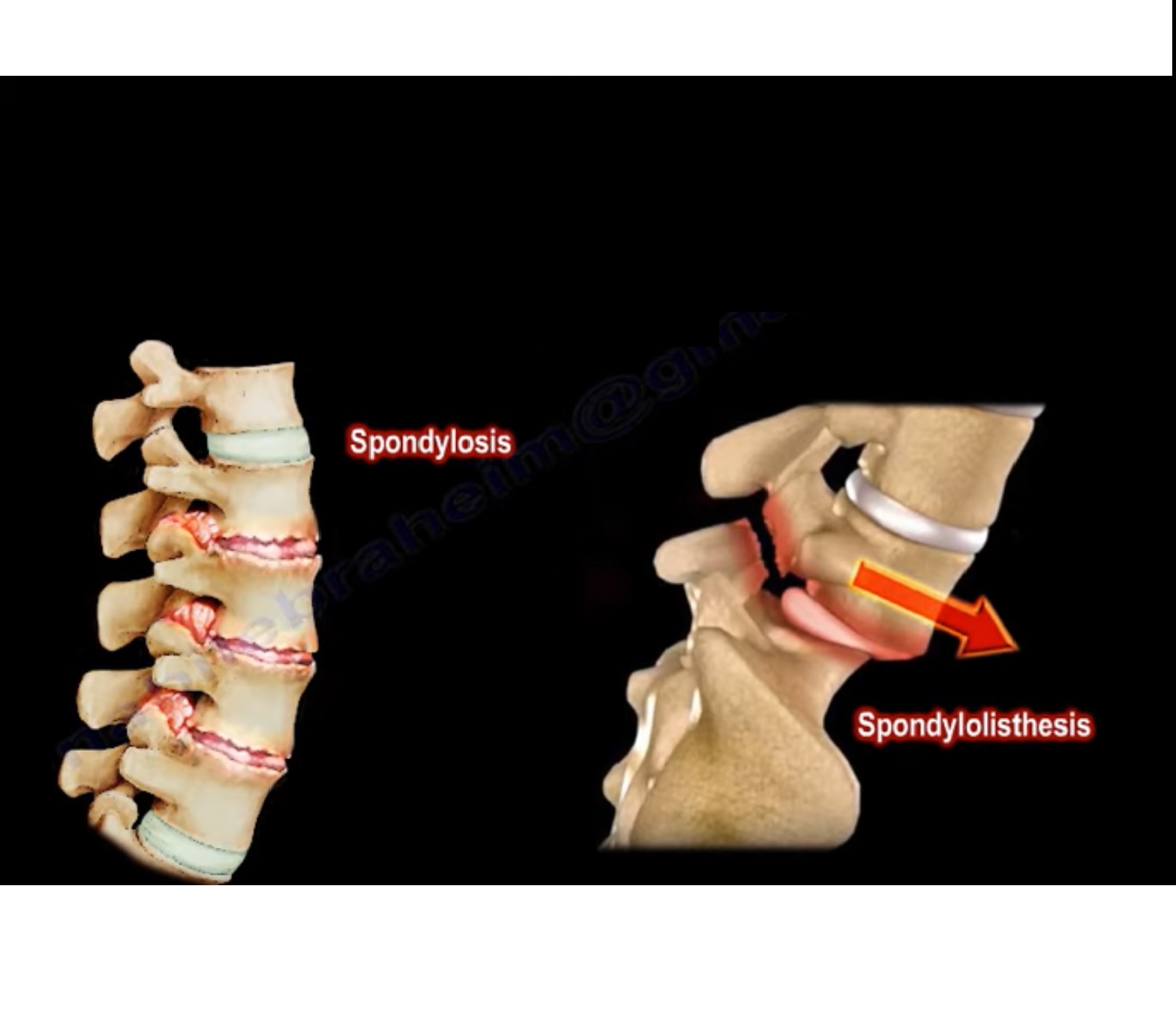
SPONDYLOLYSIS:
Bony defect in pars interarticularis is called spondylolysis.
SPONDYLOLISTHESIS:
It is the slippage of vertebrae, ie; one bone slides forward over the bone below it. It usually occurs if the stress fracture weakens the bone so much that it is unable to maintain its proper position, hence the vertebra can start to shift out of place. This slippage of vertebra compresses the nerve and narrow the spinal canal. It occur in younger age from repeated activities like gymnastics. It presents as pain with extension that progresses with time.
It is of 2 types:
a) ISTHMIC TYPE: It occurs mainly in males. L5-S1 level is the most commonly affected area. It involves the L5 nerve root with hamstring spasticity. Oblique X ray of spine shows the defect in Pars, known as the SCOTTY DOG SIGN.
b) DEGENERATIVE TYPE: Mainly occurs in middle aged females. Commonly affect the L4-L5 level. Vertebral slippage is not severe & Pars remains intact.
DEGENERATIVE SCOLIOSIS:
Degenerative changes in spinal disc & facet joints causing spine to curve & it may cause low back pain.
FAILED BACK SYNDROME:
It is the condition in which chronic back & possible leg pain occurs after spine surgery. It is mainly due to non mechanical causes like physiological factors, post operative infections or adhesions & epidural fibrosis. Or mechanical causes like inadequate surgery, recurrent disc herniation, failure of fusion &/or fixation & due to instability of spine.
For eg: If surgeon removed one facet on one side/ 50% partial facet removal on both sides, it may cause iatrogenic spondylolisthesis.
SACROILIAC JOINT DYSFUNCTION:
It is an occult cause of low back pain. It presents as pain in lower back region, usually to sides. Faber test is helpful in determining the presence of sacroiliac joint problems. The best method of diagnosis is by administering an injection into Sacroiliac joint to localise the source of pain.
Other conditions causing low back pain include Osteoporotic compression spine fracture in elderly, multiple myeloma or metastatic tumor.
Rule out other condition such as hip conditions as it may confuse the diagnosis. Also differentiate Sciatica due to disc herniation from Sciatica due to Piriformis syndrome.
90% patients with low back pain get better within 1 month. Combination of time and short period of rest with NSAIDs improve the patient condition.
Leave a Reply