Courtesy: Prof Nabil Ebraheim, University of Toledo, Ohio, USA
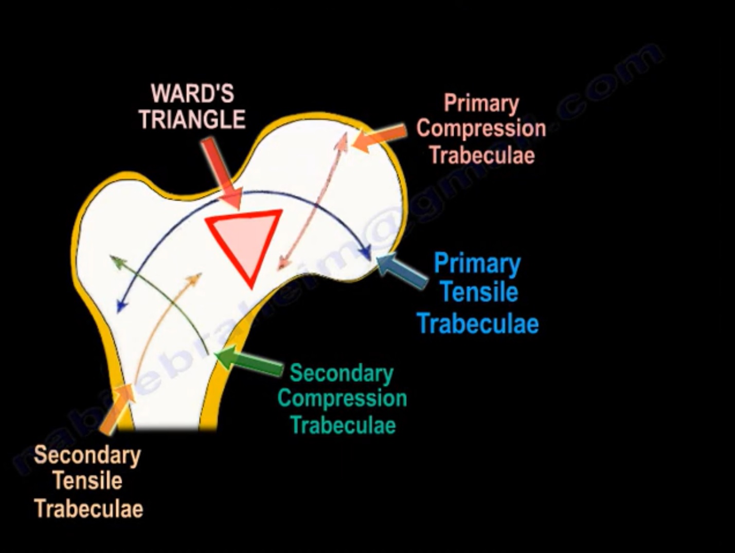
TRABECULAR PATTERN OF THE PROXIMAL FEMUR
- There are important trabecular patterns in the proximal femur.
- It occurs according to Wolff’s Law in response of the bone to stress.
- There is a primary tensile trabeculae and a primary compression trabeculae.
- There also are secondary tensile and compression trabeculae.
- In between them is what is known as the Ward triangle(very weak, empty area, not full of trabeculae). This trabecular pattern is seriously affected by osteoporosis.
- The tensile forces can create micro fractures or stress fractures at the superior neck in response to repeated loads.
- Adult bone is weak in tension.
- Stress fracture in this area will propagate and will become complete.
- Early identification and treatment by fixation of this stress fracture is important.
- Otherwise, the fracture will become complete.
- It may become displaced and may be hard to treat.
- The tip apex distance is important when treating hip fractures with compression hip screws.
- This is because the screw will be imbedded into an area where the tensile trabeculae and compression trabeculae cross each other(it is more dense area that contains more bone).
- The failure rate of the fixation is less if the tip apex distance is less than 2.5cm.
- The lateral cortex of femur is importantly because, in this area, the trabeculae present is secondary tensile trabeculae(this area is weak and can easily be affected by fractures).
- Integrity of lateral wall is important in order to achieve stable fixation and lessen the rate of revision of fixation.
- If the lateral wall integrity is compromised by a fracture or by deficiency, then the surgeon should select an intramedullary nail rather than a compression hip screw.
- In the case of hip fracture and osteoporosis, it is advisable to insert the screws posteriorly and inferiorly, because the trabecular pattern is more dense in this area.

Leave a Reply