OrthopaedicPrinciples.com

Integrating Principles and Evidence

Integrating Principles and Evidence

  • Home
  • Editorial Board
  • Our Books
    • Evidence Based Orthopaedic Principles
  • Courses
  • Exams
  • Reviews
  • Live Program
  • Contact

Anatomy of the Sartorius Muscle

Courtesy: Prof Nabil Ebraheim, University of Toledo, Ohio, USA

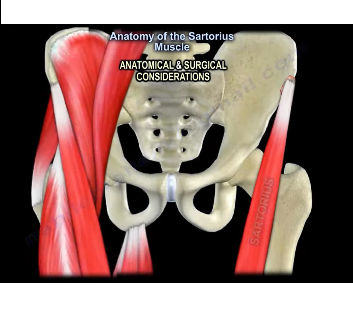
Anatomy of Sartorius
Introduction:
It is a superficial muscle and the longest muscle of the body with parallel fibres located over the anterior aspect of thigh.

Attachments:
Origin:
It takes its origin at the anterior superior iliac spine of pelvic bone.

Insertion:
It is inserted to the anteromedial upper surface of tibia, where 2 other muscle tendons (gracilis and semitendinous) also gets inserted to form pes anserine.

Course:
It crosses the upper third of thigh obliquely, downwards medially and the descend vertically towards its insertion.

Nerve supply:
The sartorius is innervated by femoral nerve.

Function:
The main actions of sartorius muscle are flexion, abduction and lateral rotation of hip and flexion of knee. Due to this action, it is also called as the “tailor’s muscle”, because it helps to position the leg in the way the tailors once did.

Anatomical significance:

  • The upper third of the sartorius forms the lateral border of femoral triangle.
  • Middle third forms the roof of adductor canal.

Clinical significance:

  • Anterior approach to hip (smith peterson approach): The neurovascular bundle passes medial to sartorius, hence it is better to approach the hip anteriorly from lateral aspect of sartorius.

Bony avulsion of sartorius tendon:

  • It occurs due to a strong sudden pull of sartorius with hip in extended position and knee in flexion. It is commonly seen in sprinters and running athletes.
  • Also when taking up iliac crest graft graft, it is always better to take it atleast 3cm away from ASIS in order to prevent avulsion.

FEMORAL TRIANGLE:

  • It is a superficial triangular space located in the anterior aspect of thigh just inferior to inguinal ligament.

Boundaries:

  • Lateral border is formed by the medial border of sartorius.
  • Medial border is formed by the medial border of adductor longus.
  • Base is formed by inguinal ligament.

Contents:
From lateral to medial the contents of this triangle are femoral nerve, femoral artery, femoral vein and deep inguinal ligaments.

ADDUCTOR CANAL(SUBSARTORIAL CANAL)

  • it is an aponeurotic tunnel in the middle third of thigh extending from apex of femoral triangle to the opening of adductor magnus (adductor hiatus).
  • The contents of this canal include femoral artery, femoral vein and saphenous nerve ( branch of femoral nerve).

PES ANSERINE BURSA:

  • Pes anserine bursa is a small fluid filled sac situated between tibia and the three tendons of sartorius, gracilis and semitendinous.
  • Innervation: the sartorius is innervated by femoral nerve, gracilis by obturator nerve and semitendinous by tibial branch of sciatic nerve.

Pes anserine bursitis ( breast stroke knee):

  • It is an inflammatory condition of medial aspect of knee at pes anserine bursa.the patient mainly presents with pain over medial aspect of knee, tenderness below the joint line on medial part of proximal tibia, and a localised swelling.
  • It usually follows a trauma or can be iatrogenically induced by total knee replacement.

Post Views: 4,836

Related Posts

  • Anatomy of the Sartorius Muscle

    Courtesy: Prof Nabile Ebraheim, University of Toledo, Ohio, USA Origin- Sartorius muscle arises from the…

  • Anatomy of Plantaris Muscle

    Courtesy: Prof Nabil Ebraheim, University of Toledo, Ohio, USA

  • Popliteus Muscle

    Courtesy : Prof Nabile Ebraheim, University of Toledo, Ohio, USA Popliteus Muscle A triangular muscle…

Leave a Reply Cancel reply

Your email address will not be published. Required fields are marked *

Follow Us

instagram slideshare

Categories

  • -Applied Anatomy
  • -Approaches
  • -Basic Sciences
  • -Cartilage & Meniscus
  • -Classifications
  • -Examination
  • -Foot and Ankle
  • -Foot and Ankle Trauma
  • -FRCS(Tr and Orth) tutorials
  • -Gait
  • -Hand and Wrist
  • -Hand and Wrist Trauma
  • -Hand Infections
  • -Hip and Knee
  • -Hip Preservation
  • -Infections
  • -Joint Reconstruction
  • -Knee Arthroplasty
  • -Knee Preservation
  • -Metabolic Disorders
  • -Oncology
  • -OrthoBiologics
  • -OrthoPlastic
  • -Paediatric Orthopaedics
  • -Paediatric Trauma
  • -Patellofemoral Joint
  • -Pelvis
  • -Peripheral Nerves
  • -Principles
  • -Principles of Surgery
  • -Radiology
  • -Rheumatology
  • -Shoulder and Elbow
  • -Shoulder and Elbow Arthroplasty
  • -Spine Deformity
  • -Spine Oncology
  • -Spine Trauma
  • -Spine, Pelvis & Neurology
  • -Sports Ankle and Foot
  • -Sports Elbow
  • -Sports Knee
  • -Sports Medicine
  • -Sports Medicine Hip
  • -Sports Shoulder
  • -Sports Wrist
  • -Statistics
  • -Technical Tip
  • -Technology in Orth
  • -Trauma
  • -Trauma (Upper Limb)
  • -Trauma Life Support
  • -Trauma Reconstruction
  • Book Shelf
  • Book Shelf Medical
  • Careers
  • Case Studies and Free Papers
  • DNB Ortho
  • Evidence Based Orthopaedic Principles
  • Evidence Based Orthopaedics
  • Exam Corner
  • Fellowships
  • Guest Editor
  • Guest Reviews
  • Image Quiz
  • Instructional Course Lectures
  • Journal Club
  • MCQs
  • Meetings and Courses
  • Multimedia
  • News and Blog
  • Plaster Techniques
  • Podcasts
  • Public Health
  • Rehabilitation
  • Research
  • Shorts and Reels

Book Shelf

Kendall’s Muscle Testing and Function 6th Edition

Kendall’s Muscle Testing and Function 6th Edition

By admin Leave a Comment

Get Book Kendall’s Muscles: Testing and Function, with Posture and Pain, 6th Edition, transforms this landmark Physical Therapy classic to prepare you for unparalleled clinical success in today’s practice. Timeless coverage of manual muscle testing, evaluation, and treatment meets the latest evidence-based practices, engaging imagery, and dynamic digital resources to create a powerful resource you […]

Popular Posts

  • Bone Screws in Orthopaedic Surgery
  • Silverskold Test
  • Piriformis Syndrome
  • Blood Supply of Long Bone
  • Movements of the Thumb

Recent Comments

  • RAJATABHA BISWAS on NUH Fellowship in Singapore
  • Runj on ESSKA Congress 2026
  • OT Hand Therapist on Interosseous Muscles Of The Hand
  • Badreddine on Rockwood and Green Fractures in Adults and Children- 10th Edition
  • Prof Dr P.sridhar MS Ortho,D Ortho on Rockwood and Green Fractures in Adults and Children- 10th Edition

Exam Corner

FRCS Orth Exam- Knee Arthroplasty

Courtesy: Zaid al Rab, FOunder, OrthoPass

MCQ Exam for the FRCS Orth 1

Courtesy: Zaid al Rub, Founder, OrthoPass

Postgraduate Entrance Exam Set 3

Get explanatory answers from our book

Postgraduate Entrance Exam Set 2

Get explanatory answers from our book

Main Menu

  • Orthopaedic Principles
  • Editorial Board
  • Orthopaedic Principles-A Review

Recent Posts

  • Anterior Tibial Tendon Tears
  • Endoscopic Lumbar Microdiscectomy
  • RAMP Lesion of the Knee
  • Osteochondritis Dissecans of the Knee
  • Dual Mobility Cups in Total Hip Replacement

Links

  • Join Our Editorial Board
  • Journals
  • Weblinks
  • Submit Your Conference
  • Disclaimer
Copyright@orthopaedicprinciples.com. All right rerserved.