OrthopaedicPrinciples.com

Integrating Principles and Evidence

Integrating Principles and Evidence

  • Home
  • Editorial Board
  • Our Books
    • Evidence Based Orthopaedic Principles
  • Courses
  • Exams
  • Reviews
  • Live Program
  • Contact

Perthe’s Disease for the FRCSOrth

Courtesy: Sally Hobson, Hull Royal Infirmary, Hull, UK

Perthes Disease
Definition:

  • Idiopathic osteonecrosis of capital femoral epiphysis in a growing child.

Epidemiology: –

  • Males 4x females.
  • 80% 4-9 (2-12 reported).
  • Social class 4-5.
  • Bilateral 10%:
  • Usually asymmetric.

Presentation – history

  • Limp.
  • +/- hip/knee pain.
  • Usually insidious/chronic onset.

Presentation – examination

  • Reduced ROM – ABDUCTION.
  • Ataxic/trendelenberg gait.
  • Wasting thigh/buttock.
  • FFD, limited rotation.
  • LLD:
    o Adduction contracture.
    o True shortening due to collapse CFE.

Differential Diagnosis

  • Acute presentation versus chronic post AVN hip xray.
  • Unilateral versus bilateral.
    Infection.
    Inflammatory.
    Skeletal dysplasia (MED).
    Haematological:
    o Sickle cell/thalassaemia.
    o Haemophilia.
    o Leukaemia.
    Metabolic.
    Trauma.
    Iatrogenic (DDH

Investigations

  • FBC/Inflammatory markers – exclude dd.
  • X-ray diagnosis – only miss if very early presentation.
  • Bone scan, MRI not usually required.
  • Arthrogram / EUA can be useful.

Classification

  • Catterall:
    % of head involvement 25/50/75/100.

Herrings lateral pillar classification:
o Comparison to contralateral, normal side.
o Based on the worst x-ray in the series.
A – lateral pillar preserved.
B ->50% maintained.
C – > 50 % involved

Aetiology

  • Poorly understood.
  • ?infection.
  • ?trauma.
  •  ?transient synovitis.
  •  ?clotting abnormality
  •  ?vascular changes primary or secondary to cartilage disorder.

Pathology – stages

  • Early/infarction.
  •  Intermediate/fragmentation.
  • Healing.
  • Remodelling.
  • Whole disease process over several years.

Pathology – sclerotic

Infarction:
o Early – Hypertrophy of cartilage and reduced epiphyseal height.
o Later – Sclerosis, subchondral fracture (crescent sign).

Pathology – fragmentation

  • Necrotic bone replaced by fibrocartilage.
  • Revascularisation by creeping substitution.
  • Areas of unossified cartilage stream across physis into metaphysis (cysts) – can cause growth arrest.

Pathology – healing

  • Endochondral ossification – fibrocartilage reossifies.
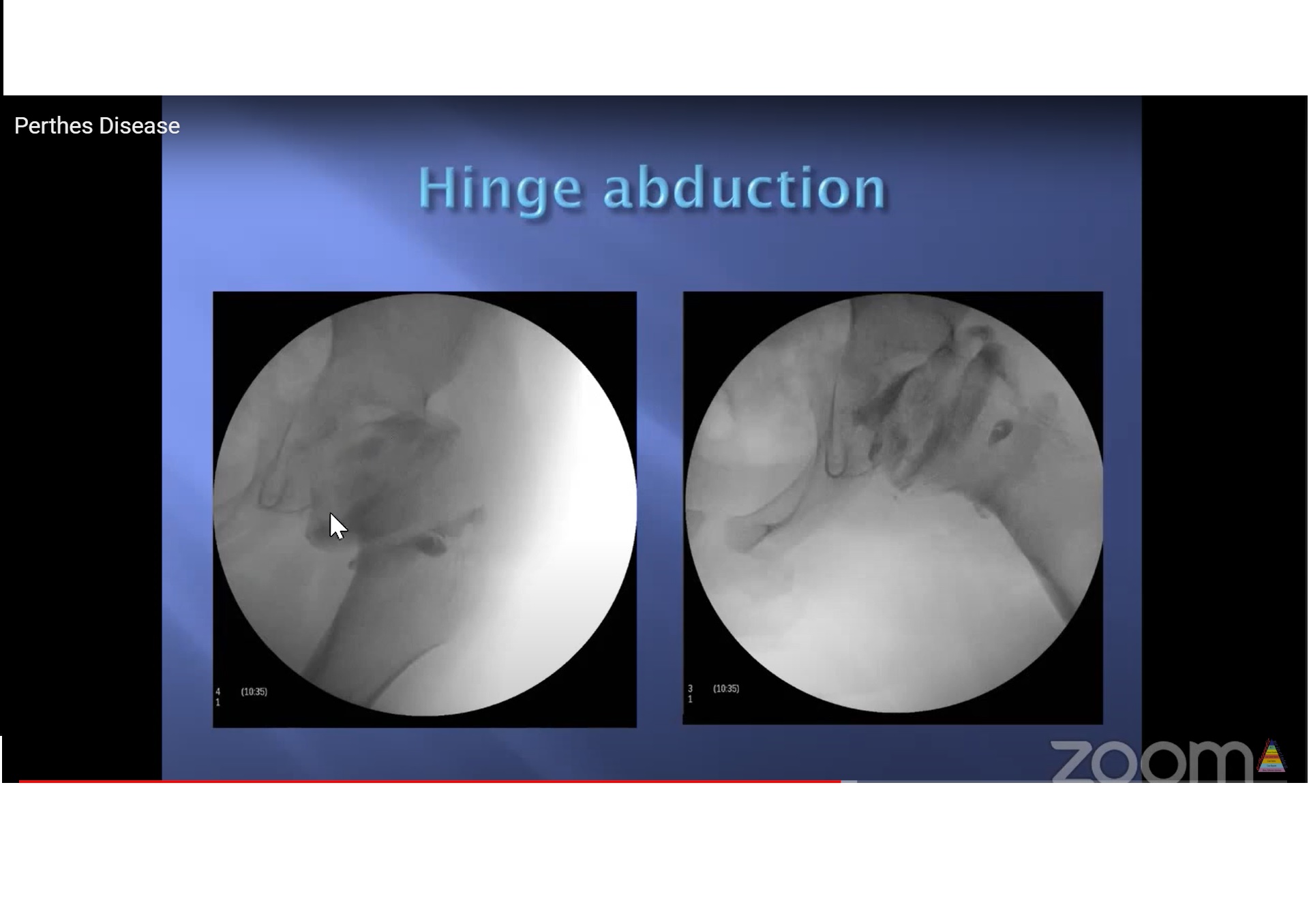
  • Last portion to form is anterosuperior epiphysis – round head becomes oval – can produce hinge abduction

Pathology – remodelling

Treatment – non-operative

  • Bed rest.
  • (Traction)
  • Activity modification/crutches.
  • Analgesia.
  • Physio to maintain ROM.
    ? (Abduction brace).
  • Herring says NONE of these affect outcome.
  • Follow up patients 3-6 monthly depending on stage in disease and if considering surgery.

Treatment – operative

  • Containment (in early/fragmentation phase):
  • EUA arthrogram to assess.
    Options:
    o Proximal femoral osteotomy.
    o Salter osteotomy.
    o Shelf osteotomy.
    ?Hip distraction.

Outcomes

Herring says:
o A all do well regardless of treatment and age.
o C all do badly regardless of treatment and age.
o B – outcome improved by surgery IF patient over 8yrs.

  • High rates of future OA/need for THR.
  • Worse with higher Stulberg grade.

Treatment – salvage

  • Valgus extension osteotomy.
  • Chiari.
  • Cheilectomy.
  • Arthroplasty

Conclusion

  • Aetiology unclear.
  • Typical presentation but remember differential diagnoses.
  • Predictable pathology.
  • Treatment and assessment of outcome controversial:
    o EUA/arthrogram to plan.
    o Maintain ROM.
    o Containment.
    o Salvage surgery for painful hinge abduction.
Post Views: 8,968

Related Posts

  • Perthe's Disease

  • Perthe's Disease- Radiology

    Courtesy: Dr Dalia Sepúlveda, Dr Lynn Staheli. GlobalHELP www.global-help.org

  • Paget's disease for the FRCSOrth

    Courtesy: Ajith Appuhamy, FRCS Tr and Orth, Srilanka

Leave a Reply Cancel reply

Your email address will not be published. Required fields are marked *

Follow Us

instagram slideshare

Categories

  • -Applied Anatomy
  • -Approaches
  • -Basic Sciences
  • -Cartilage & Meniscus
  • -Classifications
  • -Examination
  • -Foot and Ankle
  • -Foot and Ankle Trauma
  • -FRCS(Tr and Orth) tutorials
  • -Gait
  • -Hand and Wrist
  • -Hand and Wrist Trauma
  • -Hand Infections
  • -Hip and Knee
  • -Hip Preservation
  • -Infections
  • -Joint Reconstruction
  • -Knee Arthroplasty
  • -Knee Preservation
  • -Metabolic Disorders
  • -Oncology
  • -OrthoBiologics
  • -OrthoPlastic
  • -Paediatric Orthopaedics
  • -Paediatric Trauma
  • -Patellofemoral Joint
  • -Pelvis
  • -Peripheral Nerves
  • -Principles
  • -Principles of Surgery
  • -Radiology
  • -Rheumatology
  • -Shoulder and Elbow
  • -Shoulder and Elbow Arthroplasty
  • -Spine Deformity
  • -Spine Oncology
  • -Spine Trauma
  • -Spine, Pelvis & Neurology
  • -Sports Ankle and Foot
  • -Sports Elbow
  • -Sports Knee
  • -Sports Medicine
  • -Sports Medicine Hip
  • -Sports Shoulder
  • -Sports Wrist
  • -Statistics
  • -Technical Tip
  • -Technology in Orth
  • -Trauma
  • -Trauma (Upper Limb)
  • -Trauma Life Support
  • -Trauma Reconstruction
  • Book Shelf
  • Book Shelf Medical
  • Careers
  • Case Studies and Free Papers
  • DNB Ortho
  • Evidence Based Orthopaedic Principles
  • Evidence Based Orthopaedics
  • Exam Corner
  • Fellowships
  • Guest Editor
  • Guest Reviews
  • Image Quiz
  • Instructional Course Lectures
  • Journal Club
  • MCQs
  • Meetings and Courses
  • Multimedia
  • News and Blog
  • Plaster Techniques
  • Podcasts
  • Public Health
  • Rehabilitation
  • Research
  • Shorts and Reels

Book Shelf

Kendall’s Muscle Testing and Function 6th Edition

Kendall’s Muscle Testing and Function 6th Edition

By admin Leave a Comment

Get Book Kendall’s Muscles: Testing and Function, with Posture and Pain, 6th Edition, transforms this landmark Physical Therapy classic to prepare you for unparalleled clinical success in today’s practice. Timeless coverage of manual muscle testing, evaluation, and treatment meets the latest evidence-based practices, engaging imagery, and dynamic digital resources to create a powerful resource you […]

Popular Posts

  • Bone Screws in Orthopaedic Surgery
  • Silverskold Test
  • Piriformis Syndrome
  • Blood Supply of Long Bone
  • Movements of the Thumb

Recent Comments

  • RAJATABHA BISWAS on NUH Fellowship in Singapore
  • Runj on ESSKA Congress 2026
  • OT Hand Therapist on Interosseous Muscles Of The Hand
  • Badreddine on Rockwood and Green Fractures in Adults and Children- 10th Edition
  • Prof Dr P.sridhar MS Ortho,D Ortho on Rockwood and Green Fractures in Adults and Children- 10th Edition

Exam Corner

FRCS Orth Exam- Knee Arthroplasty

Courtesy: Zaid al Rab, FOunder, OrthoPass

MCQ Exam for the FRCS Orth 1

Courtesy: Zaid al Rub, Founder, OrthoPass

Postgraduate Entrance Exam Set 3

Get explanatory answers from our book

Postgraduate Entrance Exam Set 2

Get explanatory answers from our book

Main Menu

  • Orthopaedic Principles
  • Editorial Board
  • Orthopaedic Principles-A Review

Recent Posts

  • Anterior Tibial Tendon Tears
  • Endoscopic Lumbar Microdiscectomy
  • RAMP Lesion of the Knee
  • Osteochondritis Dissecans of the Knee
  • Dual Mobility Cups in Total Hip Replacement

Links

  • Join Our Editorial Board
  • Journals
  • Weblinks
  • Submit Your Conference
  • Disclaimer
Copyright@orthopaedicprinciples.com. All right rerserved.