OrthopaedicPrinciples.com

Integrating Principles and Evidence

Integrating Principles and Evidence

  • Home
  • Editorial Board
  • Our Books
    • Evidence Based Orthopaedic Principles
  • Courses
  • Exams
  • Reviews
  • Live Program
  • Contact

Blood Supply of Long Bone

Courtesy: Sameer Qureshi, Consultant Orthopaedic Surgeon, Ujjain, MP

BLOOD SUPPLY OF THE LONG BONE

BASIC ANATOMY OF THE LONG BONE

The elongated central part of the long bone is called the diaphysis. The enlarged area of the bone at the ends is called the epiphysis and the intermediate bone segment between the two is called the metaphysic s. The articular ends of the epiphyseal surface is covered by articular cartilage .The rest of the bone is covered by tough connective tissue called the periosteum. The nutrient foramen is an oblique canal usually situated in the diaphysis of the long bone.

ARTERIAL SUPPLY OF THE LONG BONE

Blood supply of the long bone accounts for 5-10% of the cardiac output. A typical long bone receives blood supply from various sources. They are the Nutrient arteries , Epiphyseal arteries , Metaphyseal arteries and periosteal arteries .
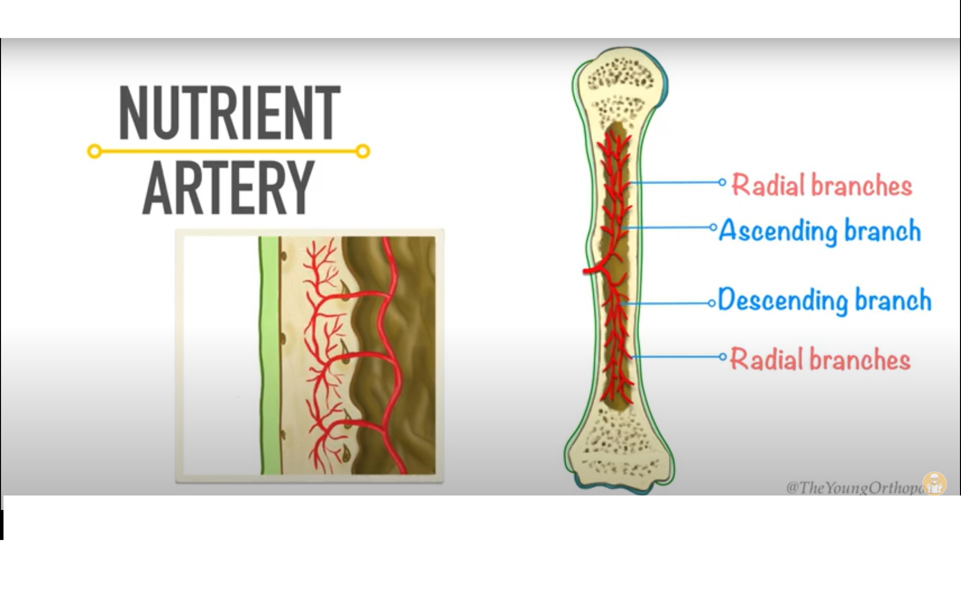
THE NUTRIENT ARTERY

The nutrient artery supplies directly from major systemic arteries. It enters the long bone through the nutrient foramen. It then divides into ascending and descending branches. These branches gives of smaller parallel arteries called the radial branches .These branches supply the bone marrow and inner third of the compact bone of the diaphysis. The ascending and descending branches at the metaphysis divides into smaller spiral branches which anastomoses with the metaphyseal and epiphyseal arteries.

THE METAPHYSEAL ARTERY

Metaphyseal arteries arising from the anastomosis around the joint enters the metaphysis at the margin of the capsule attachment. These anastomose with the spiral arteries making the metaphysic the most vascular area of the long bone.

THE EPIPHYSEAL ARTERY

Epiphyseal arteries are derived from periarticular vascular arcades. The epiphysis has openings that allows arteries to go in and out. In children the epiphyseal arteries are separated from the metaphyseal arteries due to the presence of an epiphyseal plate. In adults the epiphysis and metaphysis is fused together following the arrest of growth plate. Here the epiphyseal arteries freely anastomose with metaphyseal and nutrient arteries.

When epiphyseal cartilage and articular cartilage are continuous, the epiphyseal artery pierces the epiphyseal cartilage and supplies the epiphysis. If these arteries are damaged in epiphyseal separation, avascular necrosis may occur. In other bones where the epiphyseal cartilage is not continuous with the articular cartilage, the epiphyseal vessels enters the bone without piercing the growth plate. This helps in preventing avascular necrosis on epiphyseal separation.

THE PERIOSTEAL ARTERY

The periosteum has rich blood supply from the blood vessels that anastomose beneath the periosteum. Periosteal arteries act as a low pressure system and penetrate bone at the sides of attachment of the facial sheath or aponeurosis. They enter the Volksmann canal and supply roughly the outer one third of the compact bone of the diaphysis.

VENOUS DRAINAGE
Long bones drain into central venous sinus , then drains to nutrient veins ,then to periosteal vein and to emmissery veins successively.

Post Views: 73,705

Related Posts

  • Anatomy and Blood supply of Talus

    Courtesy: Prof Nabil Ebraheim, University of Toledo, Ohio, USA

  • Blood Supply of the Proximal Humerus

    Courtesy: Prof Nabil Ebraheim   Blood supply of the proximal humerus   1.      The axillary…

  • Blood Supply of Talus

Leave a Reply Cancel reply

Your email address will not be published. Required fields are marked *

Follow Us

instagram slideshare

Categories

  • -Applied Anatomy
  • -Approaches
  • -Basic Sciences
  • -Cartilage & Meniscus
  • -Classifications
  • -Examination
  • -Foot and Ankle
  • -Foot and Ankle Trauma
  • -FRCS(Tr and Orth) tutorials
  • -Gait
  • -Hand and Wrist
  • -Hand and Wrist Trauma
  • -Hand Infections
  • -Hip and Knee
  • -Hip Preservation
  • -Infections
  • -Joint Reconstruction
  • -Knee Arthroplasty
  • -Knee Preservation
  • -Metabolic Disorders
  • -Oncology
  • -OrthoBiologics
  • -OrthoPlastic
  • -Paediatric Orthopaedics
  • -Paediatric Trauma
  • -Patellofemoral Joint
  • -Pelvis
  • -Peripheral Nerves
  • -Principles
  • -Principles of Surgery
  • -Radiology
  • -Rheumatology
  • -Shoulder and Elbow
  • -Shoulder and Elbow Arthroplasty
  • -Spine Deformity
  • -Spine Oncology
  • -Spine Trauma
  • -Spine, Pelvis & Neurology
  • -Sports Ankle and Foot
  • -Sports Elbow
  • -Sports Knee
  • -Sports Medicine
  • -Sports Medicine Hip
  • -Sports Shoulder
  • -Sports Wrist
  • -Statistics
  • -Technical Tip
  • -Technology in Orth
  • -Trauma
  • -Trauma (Upper Limb)
  • -Trauma Life Support
  • -Trauma Reconstruction
  • Book Shelf
  • Book Shelf Medical
  • Careers
  • Case Studies and Free Papers
  • DNB Ortho
  • Evidence Based Orthopaedic Principles
  • Evidence Based Orthopaedics
  • Exam Corner
  • Fellowships
  • Guest Editor
  • Guest Reviews
  • Image Quiz
  • Instructional Course Lectures
  • Journal Club
  • MCQs
  • Meetings and Courses
  • Multimedia
  • News and Blog
  • Plaster Techniques
  • Podcasts
  • Public Health
  • Rehabilitation
  • Research
  • Shorts and Reels

Book Shelf

Kendall’s Muscle Testing and Function 6th Edition

Kendall’s Muscle Testing and Function 6th Edition

By admin Leave a Comment

Get Book Kendall’s Muscles: Testing and Function, with Posture and Pain, 6th Edition, transforms this landmark Physical Therapy classic to prepare you for unparalleled clinical success in today’s practice. Timeless coverage of manual muscle testing, evaluation, and treatment meets the latest evidence-based practices, engaging imagery, and dynamic digital resources to create a powerful resource you […]

Popular Posts

  • Bone Screws in Orthopaedic Surgery
  • Silverskold Test
  • Piriformis Syndrome
  • Blood Supply of Long Bone
  • Movements of the Thumb

Recent Comments

  • RAJATABHA BISWAS on NUH Fellowship in Singapore
  • Runj on ESSKA Congress 2026
  • OT Hand Therapist on Interosseous Muscles Of The Hand
  • Badreddine on Rockwood and Green Fractures in Adults and Children- 10th Edition
  • Prof Dr P.sridhar MS Ortho,D Ortho on Rockwood and Green Fractures in Adults and Children- 10th Edition

Exam Corner

FRCS Orth Exam- Knee Arthroplasty

Courtesy: Zaid al Rab, FOunder, OrthoPass

MCQ Exam for the FRCS Orth 1

Courtesy: Zaid al Rub, Founder, OrthoPass

Postgraduate Entrance Exam Set 3

Get explanatory answers from our book

Postgraduate Entrance Exam Set 2

Get explanatory answers from our book

Main Menu

  • Orthopaedic Principles
  • Editorial Board
  • Orthopaedic Principles-A Review

Recent Posts

  • Anterior Tibial Tendon Tears
  • Endoscopic Lumbar Microdiscectomy
  • RAMP Lesion of the Knee
  • Osteochondritis Dissecans of the Knee
  • Dual Mobility Cups in Total Hip Replacement

Links

  • Join Our Editorial Board
  • Journals
  • Weblinks
  • Submit Your Conference
  • Disclaimer
Copyright@orthopaedicprinciples.com. All right rerserved.