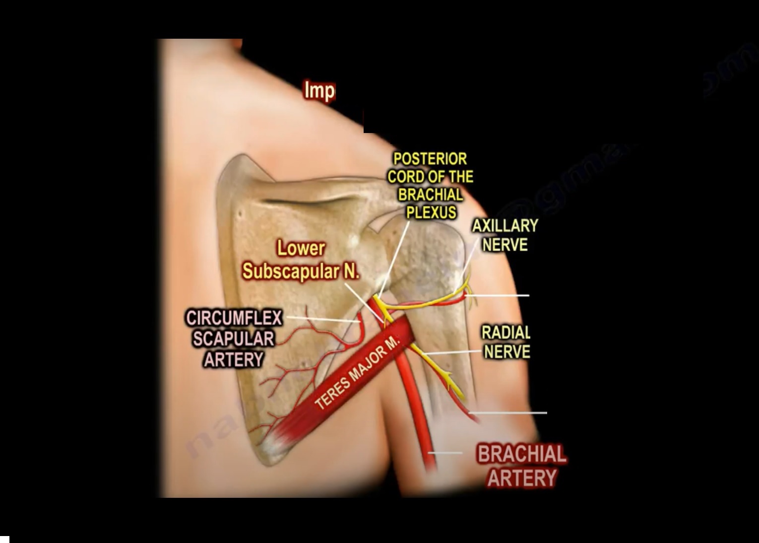
Courtesy: Prof Nabil Ebraheim, University of Toledo, Ohio, USA
Anatomy of Teres Major Muscle
- Origin- dorsal aspect of inferior angle of scapula
- Insertion- medial lip of intertubercular groove of the humerus
- One of the muscle that connect scapula to the humerus
- Teres major muscle does not attach to the capsule of glenohumeral joint
- Teres major muscle inserts in to the anterior side of proximal humerus (most medial insertion) along with latissimus dorsi pectoralis major and subscapularis muscle .
NERVE SUPPLY
- Teres major muscle is innervated by the lower subscapular nerve(C5 C6 ) of brachial plexus .
- Subscapularis muscle innervated by upper and lower subscapularis nerve (C6 C6 )
- Latissimus dorsi innervated by thoraco dorsal nerve (C6 C7 C8)
FUNCTION
- Adduction ( pull humerus towards trunk)
- Internal rotation ( turn humerus medially)
- Extension / retroversion ( pull humerus posteriorly)
ANATOMICAL STRUCTURES RELATED TO TERES MAJOR MUSCLE
QUADRANGULAR SPACE
BOUNDARIES
- Superior -teres minor muscle
- Inferior- teres major muscle
- Medial – long head of triceps
- Lateral – surgical neck of humerus
CONTENTS
- Axillary nerve
- Posterior humeral circumflex artery
Lower Triangular SPACE
BOUNDARIES
- Superior – teres major
- Medial – long head of triceps
- Lateral – shaft of humerus
CONTENTS
- Radial nerve
- Deep branch of brachial artery
Upper Triangular Space
BOUNDARIES
- Superiorly – teres minor
- Inferiorly – teres major
- Laterally- long head of triceps
CONTENTS
- Circumflex scapular artery

Leave a Reply