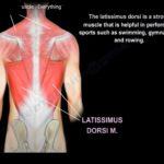
Courtesy: Prof Nabil Ebraheim, University of Toledo, Ohio, USA
Anatomy of the Latissimus Dorsi Muscle
- Latissimus dorsi muscle is anatomically is a muscle on the back but functionally is a muscle on the upper limb.
- It is the broadest muscle of the back.
- It is one of the strongest muscle and is used in sports activities like swimming, gymnastics and rowing
Origin:
1. Iliac crest
2. Thoracolumbar fascia
3. Inferior processes of inferior vertebrae (T7,T8,T9,T10,T11 and T12)
4.inferior 3 or 4 ribs
Insertion:
- inter-tubercular groove in front of the humerus.
- It lies in between the 2 muscle teres major on the medial aspect and pectorals major on the lateral aspect. All this 3 muscles lies below the insertion of. subscapularis muscle.
- Biceps tendon lie in the intertubercular groove .
Function:
- Adduction, medial rotation and extension of the arm( action at the level of shoulder joint)
- It’s action is similar to the action of teres major muscle.
- It is a synergistic muscle
Innervation:
- Thoracodorsal nerve which arises from the posterior cord
Clinical importance
1. Free flap: used as a free flap in open fracture of distal one third of tibia
2. It is used in massive rotator cuff tear ; as Latissimus dorsi tendon transfer; carried out in young adult provided the patient has intact subscapularis function
3. Latissimus dorsi muscle tear is rare and usually present with pain(athletes); occur due to eccentric overload
Treatment of Latissimus dorsi muscle tear:
1.Rest
2.physiotherapy
3. surgery: If tendon repair is to be done then it has to be carried out earlier because of chance of muscle scarring and fibrosis
Reconstruction: is the second modality , carried out in later cases

Leave a Reply