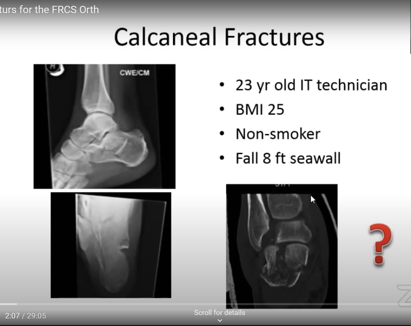
Courtesy: Rajesh Kakwani, FRCS Ortho Examiner., Foot and Ankle Surgeon, Northumbria HealthCare, UK
CALCANEAL FRACTURES
ANATOMY
- Partly articulate, Superior surface is the posterior articular facet which is the subtalar joint .
- Anteriorly it articulates with the calcaneocuboid joint and it also has the middle facet of the subtalar joint.
- The lateral surface of the calcaneum is flat and is generally targeted for fixation devices in cases of calcaneal fracture and
- The medial side has the sustendaculum tali under which past the flexor tendons and the neurovascular bundle.
- the blood supply of the calcaneal predominantly comes to the perforating branches on the medial side of the calcaneal branches of the posterior tibial artery.
- Careful while approaching the calcaneum medially – common in open fractures where the sustentaculum tali pushes through the skin and creates open fracture
- sinus tarsi region: which is between the posterior and middle articular facets of the calcaneum where there is branch of the artery of the tarsal canal and the sinus tarsi which is the main blood supply to talus.
ANGLES
1)BOHLER’S ANGLE : created by the line joining the superior aspect of the posterior calcaneal process and the superior aspect of the posture facet of the subtalar joint and the line joining between the anterior process of the calcaneum and the posterior facet. Usually 20-40 degrees
2)GISSAINE ANGLE : Between the posterior facet and the line joining it to the anterior process the calcaneal. usually between 120 to 145 degrees .
EPIDEMIOLOGY
• 75% of all foot fractures
• 1-2% of all fractures
• Male labourers who sustain bilateral intra-articular fractures and have support from compensation benefits carry a poorer prognosis
– Return to work by 4 months (light duty) and back to previous employment at 6 months
if you get a scenario of a calcaneal fracture
- first of start with ATLS protocol (because to smash the calcaneal it takes high energy)
- do complete primary and secondary survey
- 10% of calcaneal fracture patients would have a simultaneous fracture in the lumbar or dorsal number spine – can be a part of the poly trauma series
- the soft tissues around the calcaneum: it may be either open fracture or having a lot of swelling bruising or blistering- all these would affect management plan
- Prognosis(How serious is a Calcaneal fracture?): the long term implications – can be associated with a long term or may be permanent pain in limb , difficulty in mobilization , the foot shape or the heel shape will always be different and accommodative footwear may needed rather than counter footwear.
- Can Bleed into the plantar fat pad (act as bubble wrap – won’t break) so can have long-term heel pain.
-can explode into multiple pieces and it is extremely difficult to make those pieces
Common mechanism is a fall from height
- where you have forces of the body weight and the counteracting forces of the ground reaction force – the heel impacts the ground this causes a shear force which leads to the primary fracture line which is generally passing through the subtalar joint
- depending on the intensity of injury or the energy inward you can get secondary fracture lines which in a DEPRESSION TYPE of a fracture would exceed just posterior to the subtalar joint subtalar facet but in front of the Tendo Achilles attachment.
- the other type of fracture commonly is the TONGUE DEPRESSION TYPE of a fracture where you can have a version of the calcaneal tuberosity.
- now if you do have a patient with a Tongue type of a fracture – give attention to the soft tissues – can be compromise of the blood supply to the soft tissues at the level of the fracture site – they may be blanched or they may be necrosed – one of the surgical emergencies and if this is not decompressed immediately then they can lead to skin necrosis over the calcaneus insertion of the achilles tendon.
CLASSIFICATION OF CALCANEAL FRACTURES
1.Sanders classification ( common)- it involves the degree of comminution of the posterior facet of the calcaneal which is seen in its broadest area in the coronal views
TYPE 1 : it is undisplaced
TYPE 2 : major two parts
TYPE 3 : 3 part fracture
TYPE 4 : four part fracture of the posterior facet
-the constant fragment is the sustentaculum talus
-the higher type of the classification indicates more articular comminution and they would -have worse prognosis because of the extent of articular damage
How to decide whether to manage calcaneal fracture conservatively or operatively?(Does a Calcaneal Fracture require Surgery?)
-the paper by Buckley , which had more than 200 patients in either arm in our randomized control trial between conservative management and operative management of calcaneal fracture it showed that there is no functional outcome difference between the two management options
the next – the UK heel trial, the article showed that there is no difference in the functional outcomes of operative or non-operative management of calcaneal fracture although the complications are much higher in patients who underwent operative intervention
there is long-term studies , showed that the operative management is not superior in the short-term but may be beneficial in the long term for calcaneal fracture
UK heel Fracture trial
- which was a randomized control trial
- pragmatic multi-centered
- there were two arms operative and non operative
- they had 22 tertiary referral hospitals included in that trial
- 151 patients divided into the two groups the operative intervention being extensive lateral approach and the non operative management option
- follow-up of 95 % they recorded no difference in the primary outcome but the complication that re operation rate was much more common in those who would have to leave the operative care
- there was a high rate of wound complication 20% in patients who undergo operative intervention with extended lateral approach
Operative intervention
-If the patient is reliable and would comply with your operative and post-operative management
-If lateral wall blowout with calcaneofibular impingement or
-if the calcaneal height is significantly shortened
-if the posterior facet is significantly depressed or
-there is significant deformity ie varus or valgus deformity of the heel with or without calcaneofibular impingement
In a young fit patient with an articular depression of the posterior facet of the subtalar joint aims of surgery
1) Restoration of heel height
2) Correction of heel varus,
3) Tuberosity fragment control,
4) Subtalar joint reconstruction
the gold standard for surgical approach to the calcaneal – Extended lateral Approach Described by Roger Atkins in 1992
it caters to the posterior peroneal artery( blood supply to that area of the skin)
Advantage: exposure and experience.
- if this approach – mark the skin where the skin color changes from dark to light and make it an L shaped incision go straight deep up to that bone and raise a subperiosteal flap
- it is important not to take less thick flap because then the skin can necrose.
- in cases of severe wound necrosis and infection you then elevate subperiosteally until you reach the peroneal tendons you reflect the peroneal tendon superiorly until you reach the posterior facet of the subtalar joint
- At the proximal and the distal end of this surgical incision sural nerve is at risk.
The other approach that is commonly used is the Sinus Tarsi Approach
- Most popular of MIS approaches
- from the tip of the lateral malleolus to the fourth metatarsal
- it gives you direct access to the posterior facet of the subtalar joint
- but have to use indirect methods to reduce the calcaneal tuberosity fragments
Arthroscopic assisted calcaneal fracture fixation
- Percutaneous Arthroscopic Calcaneal Osteosynthesis: A Minimally Invasive Technique for Displaced Intra-Articular Calcaneal Fractures.
- able to see the articular surface quite well and use that to reduce the posterior facet but using direct methods to correct the calcaneal width and height
- shown to have lesser wound complication
- there’s no difference in the radiographic outcomes between sinus tarsi approach and the calcaneal extended lateral approach – but have much lesser complication rates in the sinus tarsi approach
Timing for surgery (When is the time to operate on Calcaneal fracture?):
Now if planning to use the extended lateral approach then it is important to wait till the swelling settles and you get skin wrinkling in the lateral aspect of the heel(the wrinkle sign)
- if you are using a sinus tarsi or an arthroscopic approach it is important to intervene as soon as possible preferably within the first two to five days
- if you have to delay surgery for some reason beyond two weeks it is extremely difficult to use the sinus tarsi approach to disimpact the fracture fragments and reduce the articular surface
Waiting for around 7 to 10 days for the wrinkle sign is not required IF- there’s a tuberosity avulsion that’s impinging on the skin – need to go using a limited approach and do it early because it might go into skin necrosis
- Requirement of an Additional medial approach – if sustentaculum tali fractures which are avulsed or in cases of open fractures you would use the medial approach
- if you have to buttress the sustentaculum tali approach it is something similar to what we would use for the coronoid process of the olecranon – any form of buttress plate would be fine , especially the one with a hook which can stabilize that sustentaculum talus fragment in that case you need to be aware of the neurovascular bundle – safe is the plane between the tibialis posterior and the FHL.
can use medial approach in closed fractures if the fragment has a spike in that region which can compress my neurovascular bundle .

Leave a Reply