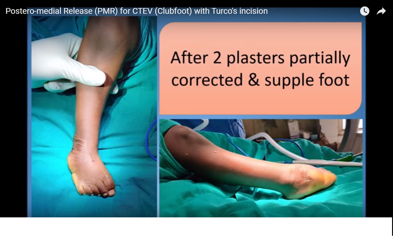
Courtesy: Premal Naik, Paed Ortho Surgeon, Ahmedabad, Gujarat, India
Surgery for CTEV is decreasing in current times and many students have never seen this surgery during their training.
PMR for CTEV is a gold standard surgery and still performed globally in patients who do not respond to Ponseti method of casting or have clubfoot along with multiple other deformities (syndromic clubfoot) or in developing countries where patients present late.
I prefer to use Turco’s posteromedial incision though many surgeons prefer Cincinnati incision. After the incision, a thick flap is raised & NV bundle is identified. Just medial to NV bundle Flexor digitorum longus (FDL) tendon and then Tibialis posterior tendons are identified. Tib post is Z lengthened followed by Tendo Achilles. In TAL medial half is detached from the heel and lateral half proximally.
Then in the foot abductor hallucis muscle is lifted from Lanciniate ligament and Talonavicular ligament. This exposes Master knot of Henry just inferior to Tib post tendon, which is released allowing easy retraction of FDL and FHL (Flexor hallucis longus)tendons. Cut distal half of Tib post is hooked out in foot after opening its sheath near navicular. Pulling on to Tib post tendon TN medial capsule is identified and released followed by lateral capsule and anterior capsule of the Subtalar joint. The release confirmed by passing the dura dissector.
This is followed by exposing and retracting FHL tendon and exposing the posterior capsule of the ankle and subtalar joints. Posterior capsule of the subtalar joint followed by ankle joint is released. Lateral capsule of the subtalar joint is also released by cutting lateral Calcaneofibular ligament. A small part of posteromedial capsule of the ankle and medial capsule of subtalar joints are released and proper release is confirmed by the dura dissector.
Correction achieved by the release is then maintained by pinning the Talonavicular joint in correct alignment. TN pinning also prevents accidental dorsiflexion of Navicular over Talus, when force is applied during postoperative casting to correct equinius. For this a k wire is passed from posteior part of Talus and brought out in centre of the Talar head. After aligning Navicular correctly over Talar head
A hemostat is then passed from proximal to distal in the foot under the Lanciniate ligament, Tib post tendon is grabbed in the tip of hemostat and is brought in the distal leg under the Lanciniate ligament to prevent bowstringing. Tib post followed by Tendo Achilles is repaired with multiple stitches. To keep gliding of TA smooth and prevent its adhesions to skin TA sheath is carefully repaired. This is a very important step for good function of TA postoperatively and leads to good recovery of ankle movements.
The tourniquet is released and hemostasis is achieved and wound is closed in layers.
Above knee POP in full correction is given for 6 weeks. Hinged Ankle Foot Orthosis (AFO) is given for 6 months full time and next 6 months night time. Ankle and subtalar range of movement exercises and TA strengthening exercises are explained to parents for home therapy program.
Children are followed up monthly for first 3 months followed by 3 monthly in the first year, six-monthly in the second year and yearly thereafter.

Leave a Reply