OrthopaedicPrinciples.com

Integrating Principles and Evidence

Integrating Principles and Evidence

  • Home
  • Editorial Board
  • Our Books
    • Evidence Based Orthopaedic Principles
  • Courses
  • Exams
  • Reviews
  • Live Program
  • Contact

Low Back Ache and Lumbar Disc Herniation

Courtesy: Prof Nabil Ebraheim, University of Toledo, Ohio, USA

The spine has bony vertebrae and intervertebral discs.

  • Inside the spinal canal lies the neural structures. The spinal cord ends at the level of T12- L1. The lower end of the spinal cord is called the Conus medullaris and from the Conus medullaris arises the Cauda equina at level L1, it consists of spinal nerves and spinal nerve rootlets
  • Low back pain is caused by various conditions of the lumbar spine including disc herniation.

Disc herniation:

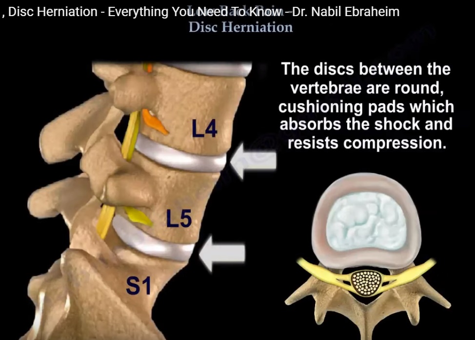
  • The lumbar spine has 5 lumbar vertebrae from L1-L5. They are attached to the sacrum at the lower end of the spine which is called the lumbosacral spine (i.e junction between lumbar spine and sacrum) L5-sacrum or tail bone.
  • The intervertebral discs between the vertebrae are round and act as a cushioning pad which absorbs the shock and resists compression.
  • The intervertebral disc is composed of 2 layers:
    1. Nucleus pulposus- it is the inner layer which is a soft gelatinous tissue.
    2. Annulus fibrosus- it is the outer layer which is strong and thick.
  • Behind the disc lies the spinal nerve root and the cauda equina. A lumbar or lumbosacral herniation could affect the nerve roots.
  • Majority cases of disc herniation is at the disc levels of L4-L5 and L5-S1.
  • The typical herniation of the L4-L5 disc level will affect the L5 nerve root. Likewise herniation of L5-S1 will affect the S1 nerve root.

Types:

1. Protrusion/bulge: It is a bulging disc with intact annular and posterior longitudinal ligament fibres.
2. Disc herniation: It is the disruption of annular fibres with tail of disc material extending into the disc space either partially or totally.
3. Sequestration: Free fragment without tail extending into the disc space. Fragment may be reabsorbed spontaneously.

Locations of disc herniation :

The most common location of disc herniation is
1. Posterolateral: It affects one nerve root (the lower one). For example- L4-L5 posterolateral disc herniation, affects the L5 nerve root.
2. Foramina: Occurs in about 8-10% of cases. It involves the exiting nerve root. For example- L4-L5 foraminal disc herniation will affect the L4 nerve root( nerve root that exits).
3. Central: It is a rare condition involving multiple nerve roots, cauda equina, predominantly causes low back pain more than leg pain. May cause bladder and bowel incontinence. It requires urgent diagnosis and surgical treatment.

Early diagnosis of central disc herniation and cauda equina syndrome is challenging. The initial signs and symptoms are visually subtle. It is important to suspect this condition and ask if the patient is having any bowel and bladder symptoms.

Do per rectal digital examination on the patient, test the perianal sensation and get emergency MRI if you suspect it.

Treatment:

  • Usually emergency surgery.
  • Timing of the surgery decides the outcome.
  • Early surgery has the best prognosis for recovery of the bladder and bowel function.

DISCOGENIC BACK PAIN – INTERNAL DISC DISRUPTION:

  • It is an early disc degeneration.
  • The patient will have annular tears.
  • Pain is worse with flexion/sitting and the pain is slightly better with extension. Forward flexion is limited on examination and the patient does not have radicular symptoms.

Post Views: 2,445

Related Posts

  • Acute Low Back Ache and Lumbar Disc Herniation

    Courtesy: Prof Nabil Ebraheim, University of Toledo, Ohio, USA

  • Lumbar Disc Herniation

    Part 1 Part 2 Courtesy: Courtesy: Douglas Gillard, BS, DC, Spine Researcher http://chirogeek.com/

  • Lumbar disc Herniation Basics

    Courtesy : Prof Nabile Ebraheim, Chairman of Orthopaedic Surgery, University of Toledo, Ohio, United states

Leave a Reply Cancel reply

Your email address will not be published. Required fields are marked *

Follow Us

instagram slideshare

Categories

  • -Applied Anatomy
  • -Approaches
  • -Basic Sciences
  • -Cartilage & Meniscus
  • -Classifications
  • -Examination
  • -Foot and Ankle
  • -Foot and Ankle Trauma
  • -FRCS(Tr and Orth) tutorials
  • -Gait
  • -Hand and Wrist
  • -Hand and Wrist Trauma
  • -Hand Infections
  • -Hip and Knee
  • -Hip Preservation
  • -Infections
  • -Joint Reconstruction
  • -Knee Arthroplasty
  • -Knee Preservation
  • -Metabolic Disorders
  • -Oncology
  • -OrthoBiologics
  • -OrthoPlastic
  • -Paediatric Orthopaedics
  • -Paediatric Trauma
  • -Patellofemoral Joint
  • -Pelvis
  • -Peripheral Nerves
  • -Principles
  • -Principles of Surgery
  • -Radiology
  • -Rheumatology
  • -Shoulder and Elbow
  • -Shoulder and Elbow Arthroplasty
  • -Spine Deformity
  • -Spine Oncology
  • -Spine Trauma
  • -Spine, Pelvis & Neurology
  • -Sports Ankle and Foot
  • -Sports Elbow
  • -Sports Knee
  • -Sports Medicine
  • -Sports Medicine Hip
  • -Sports Shoulder
  • -Sports Wrist
  • -Statistics
  • -Technical Tip
  • -Technology in Orth
  • -Trauma
  • -Trauma (Upper Limb)
  • -Trauma Life Support
  • -Trauma Reconstruction
  • Book Shelf
  • Book Shelf Medical
  • Careers
  • Case Studies and Free Papers
  • DNB Ortho
  • Evidence Based Orthopaedic Principles
  • Evidence Based Orthopaedics
  • Exam Corner
  • Fellowships
  • Guest Editor
  • Guest Reviews
  • Image Quiz
  • Instructional Course Lectures
  • Journal Club
  • MCQs
  • Meetings and Courses
  • Multimedia
  • News and Blog
  • Plaster Techniques
  • Podcasts
  • Public Health
  • Rehabilitation
  • Research
  • Shorts and Reels

Book Shelf

Kendall’s Muscle Testing and Function 6th Edition

Kendall’s Muscle Testing and Function 6th Edition

By admin Leave a Comment

Get Book Kendall’s Muscles: Testing and Function, with Posture and Pain, 6th Edition, transforms this landmark Physical Therapy classic to prepare you for unparalleled clinical success in today’s practice. Timeless coverage of manual muscle testing, evaluation, and treatment meets the latest evidence-based practices, engaging imagery, and dynamic digital resources to create a powerful resource you […]

Popular Posts

  • Bone Screws in Orthopaedic Surgery
  • Silverskold Test
  • Piriformis Syndrome
  • Blood Supply of Long Bone
  • Movements of the Thumb

Recent Comments

  • RAJATABHA BISWAS on NUH Fellowship in Singapore
  • Runj on ESSKA Congress 2026
  • OT Hand Therapist on Interosseous Muscles Of The Hand
  • Badreddine on Rockwood and Green Fractures in Adults and Children- 10th Edition
  • Prof Dr P.sridhar MS Ortho,D Ortho on Rockwood and Green Fractures in Adults and Children- 10th Edition

Exam Corner

FRCS Orth Exam- Knee Arthroplasty

Courtesy: Zaid al Rab, FOunder, OrthoPass

MCQ Exam for the FRCS Orth 1

Courtesy: Zaid al Rub, Founder, OrthoPass

Postgraduate Entrance Exam Set 3

Get explanatory answers from our book

Postgraduate Entrance Exam Set 2

Get explanatory answers from our book

Main Menu

  • Orthopaedic Principles
  • Editorial Board
  • Orthopaedic Principles-A Review

Recent Posts

  • Anterior Tibial Tendon Tears
  • Endoscopic Lumbar Microdiscectomy
  • RAMP Lesion of the Knee
  • Osteochondritis Dissecans of the Knee
  • Dual Mobility Cups in Total Hip Replacement

Links

  • Join Our Editorial Board
  • Journals
  • Weblinks
  • Submit Your Conference
  • Disclaimer
Copyright@orthopaedicprinciples.com. All right rerserved.