Courtesy: Prof Nabil Ebraheim, University of Toledo, Ohio, USA
ANTERIOR INTEROSSEOUS NERVE
Anterior interosseous nerve is a motor nerve which arises from the median nerve of brachial plexus about 4-6 cm distal to the elbow( approximately 1/3 of the way down the forearm) .
Anterior interosseous nerve exits from the anterolateral aspect of the median nerve and runs between the radius and the ulna on the interosseous membrane between and below the muscles of the flexor digitorum profundus and the flexor pollicis longus. It innervates three muscles in the forearm: flexor pollicis longus, lateral half of flexor digitorum profundus (index and long finger) and pronator quadratus.
Anterior interosseous nerve passes dorsal to the pronator quadratus muscle with the anterior interosseous artery and provides innervation to the volar wrist capsule. It’s terminal branches innervates the carpal joint capsule.
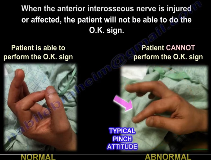
The integrity of anterior interosseous nerve is tested by performing the ‘OK sign’ or ‘circle sign’or “Kiloh Nevin sign” which can be done by asking the patient to touch the tips of the index finger and thumb together. The integrity of the nerve allows the flexion of the distal phalanx of the thumb and the index finger.
When anterior interosseous nerve is injured, the patient will not be able to bring together the distal phalanx of the thumb and the index finger. Treatment is usually observation with EMG testing. In anterior interosseous nerve entrapment, the median nerve conduction study will be normal, however the needle EMG of the anterior interosseous innervated muscles will be abnormal. Exploration and release of the nerve can be considered if no improvement in the nerve function is seen in 4-6 months.
Anterior interosseous nerve injury should be differentiated from high median nerve injury. If the patient is unable to do the ‘OK sign’ and is associated with the presence of sensory symptoms ,it indicates a high median nerve injury.
‘OK sign’ is different from Froment’s sign that seen in ulnar nerve injury. In Froment’s sign, when pinching a piece of paper between the thumb and the index finger, the thumb IP joint will flex if the adductor pollicis muscle is weak. This muscle is innervated by ulnar nerve and with ulnar nerve injury, the adductor pollicis muscle will becomes weak and the patient cannot adduct the thumb. The flexor pollicis longus muscle substitutes this movement by flexing the thumb.
Benediction sign is an another sign that can occur with anterior interosseous nerve injury. This can be performed by asking the patient to make a fist, the 1st and 2nd fingers will have difficulty in flexing, while the other digits will flex in anterior interosseous nerve injury. This position of the hand is similar to the position taken during a hand blessing. This sign should be differentiated from ulnar claw hand which occurs due to damage to the ulnar nerve and is seen when attempting to extend all the digits. In ulnar claw hand, the 4th and 5th fingers take a flexed position.
The Martin-Gruber anastomosis occurs through a communicating nerve branch between the median and the ulnar nerve in the forearm. The connection carries motor nerve fibres and it can go from the anterior interosseous nerve to the ulnar nerve. The Martin-Gruber anastomosis nerve fibres are most often of an efferent nature, contributing to the motor innervation of the hand through ulnar nerve. The axons will leave the median nerve or the anterior interosseous nerve, crossing through the forearm to join the main trunk of the ulnar nerve, innervating the intrinsic muscles of the hand. It can be confusing clinically and also on EMG. If the communicating nerve arises from the anterior interosseous nerve, the patient with anterior interosseous nerve palsy may present with intrinsic muscle weakness normally supplied by the ulnar nerve.

Leave a Reply