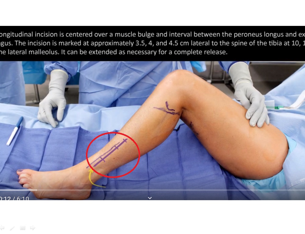
Superficial Peroneal Nerve Release in the Lower Leg – Standard
Authors: Mackinnon SE1, Yee A1
AUTHOR INFORMATION
1Division of Plastic and Reconstructive Surgery, Washington University, St. Louis, Missouri
DISCLOSURE
No authors have a financial interest in any of the products, devices, or drugs mentioned in this production or publication.
ABSTRACT
Compression of the superficial peroneal nerve (SPN) is due to the superficial fascial layer that encapsulates the SPN and its distal entrapment point called the transverse crural ligament. These structures are typically the cause for numbness and pain in the territory of the SPN. Release of the SPN involves the longitudinal release of the superficial fascial layer and the transverse crural ligament. A lateral and anterior fasciotomy is also performed both longitudinally and transversely. Care is taken to look for two branches of the SPN and decompress both branches. This release is performed on patients that present with peroneal neuropathy that fail to resolve from conservative management and have symptoms that localize to the territory of the SPN. In this case, the patient presented with a complex history of neuropathic pain in the lower left leg following multiple knee surgeries over a span of many years. During examination, the patient was able to tolerate light touch related to compression-type injury rather than withdrawing from severe pain in keeping with a neurectomy-type injury; thus compression neuropathy and not neuroma injury was suspected. The scratch collapse test with ethyl chloride revealed provocation, first at the common peroneal nerve at the fibular head, then second at the saphenous nerve in the thigh, and then third at the SPN. Her surgical management included the release of these three nerves. This video outlines the surgical technique for releasing the SPN in the lower leg.

Leave a Reply