OrthopaedicPrinciples.com

Integrating Principles and Evidence

Integrating Principles and Evidence

  • Home
  • Editorial Board
  • Our Books
    • Evidence Based Orthopaedic Principles
  • Courses
  • Exams
  • Reviews
  • Live Program
  • Contact

Anatomy of the Radial Nerve

  • Courtesy: Prof Nabil Ebraheim, University of Toledo, Ohio, USA

    RADIAL NERVE


  • It is a major peripheral nerve in the upper limb.
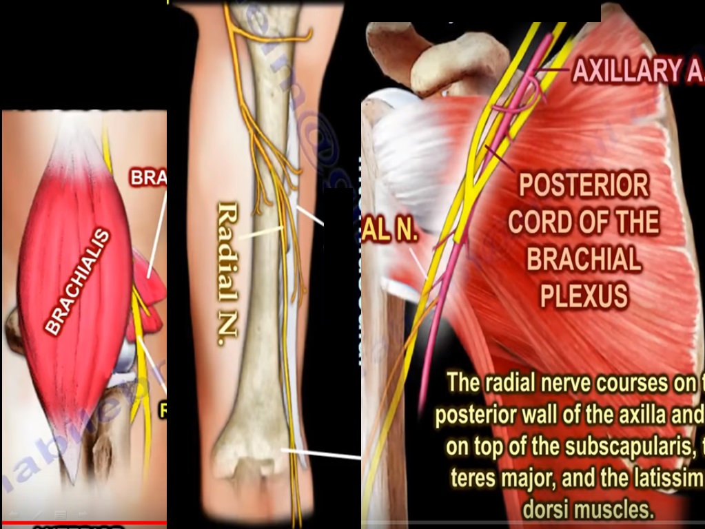
  • It arises from the posterior cord of brachial plexus and contains fibres from nerve roots C5 to T1

COURSE

  • It arises in the axilla posterior to axillary artery
  • Traverses through the posterior wall of axilla and lies on top of 3 muscles – subscapularis, teres major and latissimus dorsi
  • It gives off 3 branches in the axilla
    1.Branch to long head of triceps
    2.Branch to medial head of triceps
    3.Posterior cutaneous nerve of arm
  • Radial nerve exits the axilla inferiorly via the triangular interval with the profunda brachii artery, which is a branch of axillary artery
  • The triangular interval is bounded by teres major proximally, long head of triceps medially and humeral shaft laterally.
  • The Radial nerve then enter the upper arm between the long and medial heads of triceps and then runs through the spiral groove of humerus, which is a thin bare area of bone which lies in the upper 2/3 rd of back of humerus, between the lateral and medial heads of triceps
  • Hence the safe zones of humerus posteriorly are
    1.10 cm distal to the latetal acromion
    2.10 cm proximal to lateral epicondyle
    In the spiral groove radial nerve gives rise to 4 branches
    1.Branch to medial head of triceps
    2.Branch to lateral head of triceps
    3.Lower lateral cutaneous nerve of arm
    4.Posterior cutaneous nerve of forearm
  • Then it passes through the lateral intermuscular septum and enters the anterior compartment of the arm atleast 7.5 cm above the elbow joint
  • Anteriorly the nerve runs between brachialis and brachioradialis, anterior to the lateral epicondyle and this is the site for exposure of radial nerve anteriorly.
  • It then gives off 3 branches to supply
    1.Lateral part of brachialis
    2.Brachioradialis
    3.Extensor carpi radialis brevis
  • Radial nerve is vulnerable to injury below the spiral groove when there is fracture to distal third of humeral shaft which then leads to wrist drop
  • At the level of lateral epicondyle the nerve divides into deep and superficial braches.
  • After giving supply to the anconeus muscle the deep branch or the posterior interosseous nerve enters the extensor compartment of forearm between the two heads of supinator muscles.
  • The area in which the posterior interosseous nerve passes through is called the Arcade of Frohse and is often a site for entrapment of the nerve
  • The posterior interosseous nerve, which is a pure motor nerve, then supplies the muscles of the forearm- Extensor indicis, Extensor pollicis longus, Extensor pollicis brevis and Abductor pollicis longus
  • Injury to the posterior interosseous nerve causes inability to extend the fingers or hitchike the thumb
    And during the recovery, extensor digitorum is the 1st one to recover and extensor indicis is the last one to recover
  • The superficial radial nerve runs deep to the brachioradialis muscle and continues until about 5 cm above the wrist where it emerges from underneath brachioradialis piercing the deep fascia and lying between the brachioradialis and the extensor carpi radialis longus and then descends towards the anatomical snuff box.
  • It also gives sensory supply to majority of dorsum of hand

Wartenbergs syndrome

  • Entrapment of superficial branch of radial nerve above wrist and is manifested as pain at about 8 cm proximal to radial styloid
Post Views: 9,557

Related Posts

  • Ulnar Nerve Anatomy

    Courtesy: Prof Nabil Ebraheim, University of Toledo, Ohio, USA

  • Radial Nerve Course

    Courtesy: Prof Nabil Ebraheim, University of Toledo, Ohio, USA

  • Anatomy of the Radial Nerve

    Courtesy: Courtesy: Prof Nabile Ebraheim, University of Toledo, Ohio, United States

Leave a Reply Cancel reply

Your email address will not be published. Required fields are marked *

Follow Us

instagram slideshare

Categories

  • -Applied Anatomy
  • -Approaches
  • -Basic Sciences
  • -Cartilage & Meniscus
  • -Classifications
  • -Examination
  • -Foot and Ankle
  • -Foot and Ankle Trauma
  • -FRCS(Tr and Orth) tutorials
  • -Gait
  • -Hand and Wrist
  • -Hand and Wrist Trauma
  • -Hand Infections
  • -Hip and Knee
  • -Hip Preservation
  • -Infections
  • -Joint Reconstruction
  • -Knee Arthroplasty
  • -Knee Preservation
  • -Metabolic Disorders
  • -Oncology
  • -OrthoBiologics
  • -OrthoPlastic
  • -Paediatric Orthopaedics
  • -Paediatric Trauma
  • -Patellofemoral Joint
  • -Pelvis
  • -Peripheral Nerves
  • -Principles
  • -Principles of Surgery
  • -Radiology
  • -Rheumatology
  • -Shoulder and Elbow
  • -Shoulder and Elbow Arthroplasty
  • -Spine Deformity
  • -Spine Oncology
  • -Spine Trauma
  • -Spine, Pelvis & Neurology
  • -Sports Ankle and Foot
  • -Sports Elbow
  • -Sports Knee
  • -Sports Medicine
  • -Sports Medicine Hip
  • -Sports Shoulder
  • -Sports Wrist
  • -Statistics
  • -Technical Tip
  • -Technology in Orth
  • -Trauma
  • -Trauma (Upper Limb)
  • -Trauma Life Support
  • -Trauma Reconstruction
  • Book Shelf
  • Book Shelf Medical
  • Careers
  • Case Studies and Free Papers
  • DNB Ortho
  • Evidence Based Orthopaedic Principles
  • Evidence Based Orthopaedics
  • Exam Corner
  • Fellowships
  • Guest Editor
  • Guest Reviews
  • Image Quiz
  • Instructional Course Lectures
  • Journal Club
  • MCQs
  • Meetings and Courses
  • Multimedia
  • News and Blog
  • Plaster Techniques
  • Podcasts
  • Public Health
  • Rehabilitation
  • Research
  • Shorts and Reels

Book Shelf

Kendall’s Muscle Testing and Function 6th Edition

Kendall’s Muscle Testing and Function 6th Edition

By admin Leave a Comment

Get Book Kendall’s Muscles: Testing and Function, with Posture and Pain, 6th Edition, transforms this landmark Physical Therapy classic to prepare you for unparalleled clinical success in today’s practice. Timeless coverage of manual muscle testing, evaluation, and treatment meets the latest evidence-based practices, engaging imagery, and dynamic digital resources to create a powerful resource you […]

Popular Posts

  • Bone Screws in Orthopaedic Surgery
  • Silverskold Test
  • Piriformis Syndrome
  • Blood Supply of Long Bone
  • Movements of the Thumb

Recent Comments

  • RAJATABHA BISWAS on NUH Fellowship in Singapore
  • Runj on ESSKA Congress 2026
  • OT Hand Therapist on Interosseous Muscles Of The Hand
  • Badreddine on Rockwood and Green Fractures in Adults and Children- 10th Edition
  • Prof Dr P.sridhar MS Ortho,D Ortho on Rockwood and Green Fractures in Adults and Children- 10th Edition

Exam Corner

FRCS Orth Exam- Knee Arthroplasty

Courtesy: Zaid al Rab, FOunder, OrthoPass

MCQ Exam for the FRCS Orth 1

Courtesy: Zaid al Rub, Founder, OrthoPass

Postgraduate Entrance Exam Set 3

Get explanatory answers from our book

Postgraduate Entrance Exam Set 2

Get explanatory answers from our book

Main Menu

  • Orthopaedic Principles
  • Editorial Board
  • Orthopaedic Principles-A Review

Recent Posts

  • Anterior Tibial Tendon Tears
  • Endoscopic Lumbar Microdiscectomy
  • RAMP Lesion of the Knee
  • Osteochondritis Dissecans of the Knee
  • Dual Mobility Cups in Total Hip Replacement

Links

  • Join Our Editorial Board
  • Journals
  • Weblinks
  • Submit Your Conference
  • Disclaimer
Copyright@orthopaedicprinciples.com. All right rerserved.