Courtesy: Prof Nabil Ebraheim, University of Toledo, Ohio, USA
- In general, surgical approach to the acetabulum depends on the location of the fracture, the type of fracture, and complexity of the fracture.
- Types of fractures approached posteriorly: posterior wall fracture, posterior column fracture, posterior column fracture and posterior wall fracture, and posterior wall fracture with transverse fracture.
- The ilioinguinal approach is usually good for: anterior wall fracture, anterior column fracture, associated anterior and posterior hemi-transverse fracture, and associated BOTH column fracture.
- The transverse fracture of the acetabulum is usually approached by the ilioingunal approach if the fracture is HIGH. However, in LOW transverse fractures the fracture is approached posteriorly.
- The T-shaped fracture may require two approaches, the anterior approach and the posterior approach.
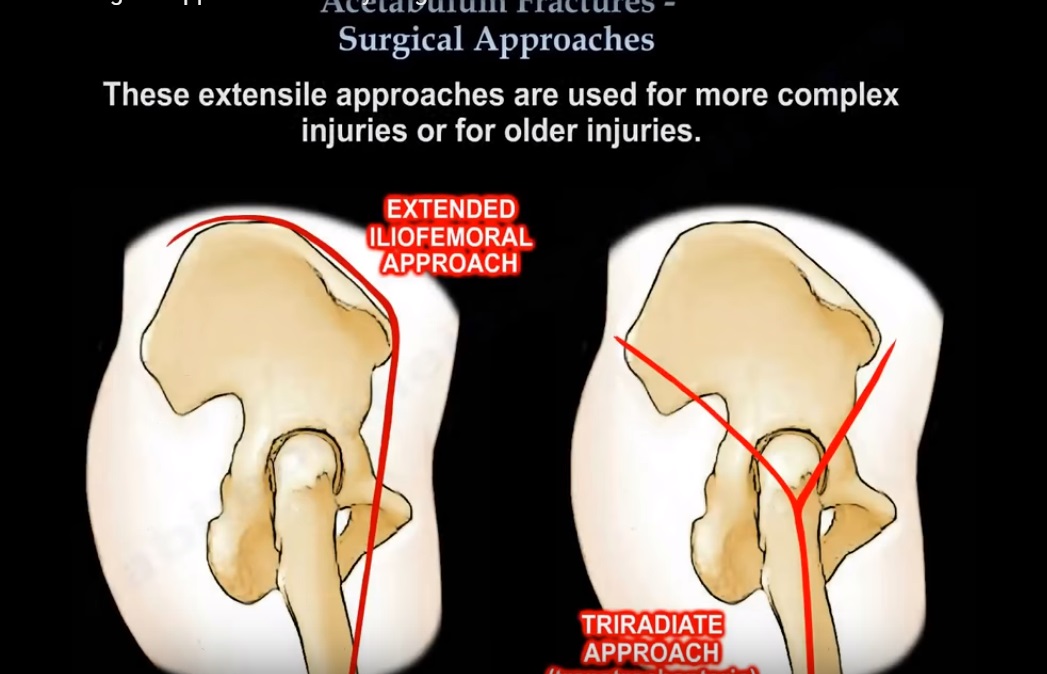
- The extensile approach can be extended iliofemoral approach or a triradiate approach.
- The extensile approaches are used for more complex injuries or for older injuries.
- The extended iliofemoral approach may cause gluteal muscle necrosis.
- The entire gluteal mass (gluteus medius and minimus) can be hanging by the superior gluteal vessels pedicle.
- The extensile approaches in general will have more myositis ossificans. Over 1/3 of them will get a severe type of myositis. You want to prevent this by giving low dose radiation (600-800 centigray of radiation) given within 72 hours of the surgery or you can give Indomethacin for 6 weeks (decreases the severity of the myositis but it may not prevent it).
- In general, most surgeons will do an anterior and posterior approach (dual approach) instead of the extensile iliofemoral approach.
- The triradiate approach is a triradiate transtrochanteric approach.
- The ilioinguinal approach is known to have a low incidence of myositis.
- Myositis is 10% in the posterior approach and 1% in the ilioinguinal approach.
- With the ilioinguinal approach, you can approach the anterior column, anterior wall lesions associated both columns fractures, high transverse fractures, and associated anterior and posterior hemi-transverse fractures. There are 3 windows of the ilioinguinal approach.
- First is the medial window, it contains the spermatic cord and the ilioinguinal nerve.
- Second is the middle window, it contains the external iliac vessels and may carry the Corona Mortis (vascular zone). The external iliac vessels are the same vessels that are seen in the anterior superior quadrant (external iliac vessels may be injured by placement of screws during total hip replacement).
- Third is the lateral window, this contains the iliopsoas muscle, the femoral nerve, and the lateral femoral cutaneous nerve of the thigh. These nerves can be injured during an ilioinguinal approach!
- The iliopectineal fascia lies between the middle window and the lateral window. You want to incise the iliopectineal fascia down to the eminence of the pelvic brim. After you do this, then you will be able to connect the true pelvis to the false pelvis.
- Three important problems with the ilioinguinal approach: lateral cutaneous nerve of the thigh, may develop a hernia if repair of the anterior abdominal wall is not good, and corona mortis.
- Corona mortis is a connection between the internal iliac branch (obturator) and the external iliac or its branch, the inferior epigastric. Its location on the superior pubic ramus is variable.
- It is about 3-7 cm from the symphysis pubis. The corona mortis is susceptible to injury in the pelvic trauma and in the pelvic surgery especially during the ilioinguinal approach. Injury to the corona mortis may lead to significant hemorrhage which may be difficult to control.
- Posterior approach is Useful in displaced posterior fractures of the acetabulum, such as posterior wall fracture, posterior column fracture, in associated fractures which have a larger posterior wall fracture. Posterior approach is also useful in some transverse fractures.
- If you combine the posterior approach with an anterior incision and trochanteric osteotomy, then this can make the approach more extensile.
- Sliding trochanteric osteotomy will improve visualization at the acetabular dome and the superior acetabulum.
- Problems with the posterior approach: cannot reach the anterior displaced fractures adequately and injury to the sciatic nerve may occur.
- When you do posterior approach to the acetabulum, you need to bend the knee and extend the hip. Need to keep the knee bent all the time, especially during traction to avoid injury to the sciatic nerve.

Leave a Reply