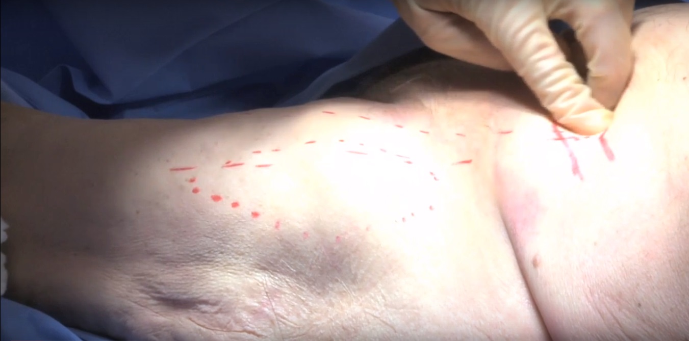
Courtesy: ToJo Productions, Baltimore, USA and Dr Jason Nascone, Associate Professor, University of Maryland

Smith Peterson approach*
Smith Peterson approach to the hip :
Indications :
Femoral head fractures
Femoral neck fractures
Select Anterior column fractures
Select anterior wall fractures
Modified Iliofemoral approach/modified smith Peterson approach :
Indications:
Select anterior wall fractures
Select anterior column fractures
Periacetabular osteotomies,PAO
Landmarks include the anterior superior iliac spine showing the sartorius tendon, the tensor fascia lata,palpation of the muscle belly of tensor fascia.
Incision is anterior over the hip, it is carried down to the fascia of the tensor fascia lata. Once the muscle belly is identified with palpation and direct visualisation, the fascia should be incised and the muscle of the tensor fascia should be mobilised in the lateral direction. This maintains the anterior and medial fascia of the muscle belly protecting the lateral femoral cutaneous nerve. Careful dissection is performed at this level to mobilise the muscle belly in the lateral direction.
Next step would be to incise the floor of the tensor fascia lata muscle belly to obtain access to the next interval, to provide access to the rectus femoris. This layer will have multiple fascial planes. The muscle belly of the rectus is identified and mobilised in the medial direction to provide exposure to the anterior hip capsule and the underlying capsule layers. The Rectus femoris tendon will be identified in a more proximal direction which has two heads ; a direct and an indirect head. Direct head attaching at the anterior inferior iliac spine and the indirect head attatching at the lateral aspect of the acetabulum. Further releasing of the tensor from the external iliac ring will facilitate a wider exposure. This approach can be extended to proximal and distal direction.
Now dissection will be performed in the medial aspect of the rectus tendon. Next step is tenotomy of the rectus tendon to provide additional visualisation of the anterior hip capsule. This may or may not be required depending on the particular needs of the approach. Distally the tendon is released and mobilised in a caudal direction. Now the fibres of the capsule layers will be mobilised. It’s a very fine layer of muscle over the hip capsule which can be mobilised in a medial direction, exposing the hip capsule anteriorly upto the level of psoas tendon.
Finger palpation will help to identify the inferior aspect of the capsule. As the dissection extends in the medial direction the bursa will be entered. This is the bursa of the iliopsoas. And proximal palpation will lead the surgeon over the pelvic brim to psoas gutter , here the psoas tendon is visualised. Further mobilisation of the psoas tendon will give the surgeon an increased exposure to the pubic root and ramus. Distally the vessels of the anterior circumflex system can be visualised and they may or maybe not be ligated depending upon the level of exposure. The system usually consist of two veins and one artery.
Now the hip is mobilised to adequately visualise where the acetabulum and the femoral head meet, to check the best location for capsulotomy.
Care must be taken while incising the capsule so as not to cut the labrum. Palpation maybe useful to delineate the capsule- labrum margin is located.
There are multiple options for capsulotomy depending on the fracture that’s being addressed. A T shaped incision or an incision along the intertrochanteric region or along the capsule labrum junction or a combination.
In this example ,capsule is performed along the intertrochanteric line and a T shaped incision will be extended towards the capsule labral junction. This will give increased exposure to the base of the neck and the proximal femur. Similarly if the capsulotomy was oriented in the other direction it would give a better exposure to the acetabulum and a more proximal aspect of femoral head.
Labrum is seen, which is ossified.
Retractors can be placed along the inferior aspect of the neck as needed. Care must be taken in positioning the retractors as it was violate the blood supply to the femoral head.
The inferior aspect of the capsule can be rotated in the inferior direction and this displays the inferior aspect of the femoral head.
This capsulotomy is reapproximated in an effort to show the change in exposure with a more proximal capsulotomy. The inferior limb along the intertrochanteric line is reapproximated , the vertical limb
Is left intact and then capsulotomy at the capsule labral junction is performed to show the difference in view and the difference in exposure.
These can be combined for an H shaped capsulotomy if needed.
The visualisation shows increased exposure to the anterior inferior femoral head.
The approach can be extended to a proximal direction to an iliofemoral approach by curving the incision along the anterior iliac crest. The raphe between the external oblique musculature and the gluteus medius musculature will be identified along the iliac crest and sharp dissection need to be performed. Muscles are then released from the external aspect of the ilia ring by sharp dissection. This provides increased exposure to the external aspect of the ilium and the supraacetabular region.
Finger palpation along the inter spinous notch between the ASIS and AIIS provide access to the internal aspect of acetabulum. The dissection can also be extended to the internal aspect of the iliac fossa with release of the external oblique musculature from the ilium. Further dissection could be performed in the internal aspect providing exposure to the internal aspect of iliac fossa, this would be a modification of the iliofemoral approach. In this situation an ASIS osteotomy is performed in an effort to mobilise the sartorius and inguinal ligament in a medial direction to provide increased exposure to internal aspect of acetabulum. This is later reattached with small fragment screw fixation.
With mobilisation medially, the remaining soft tissue from the AIIS is released. Now full exposure to supra acetabular region on the internal aspect of the ilium is obtained down to the level of the pubic root.
Leave a Reply