Courtesy: Prof Nabil Ebraheim,
University of Toledo, Ohio, USA
Intertrochanteric fracture
- It occurs in the region between greater trochanter and lesser trochanter
- There is primary tensile trabeculae and compressive trabeculae and secondary tensile trabeculae and compressible trabeculae
- In between this there is triangle called wards triangle which is responsible for this stress fracture
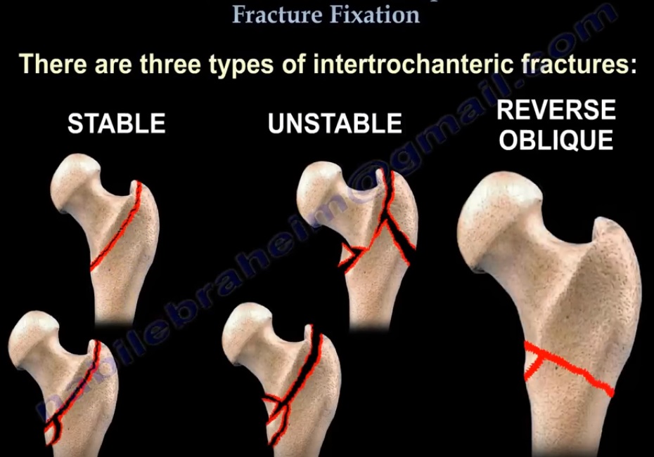
- There are three type of intertrochanteric fracture – stable, unstable, reverse oblique. Occasionally IT fracture extend to form a subtrochanteric fracture
Stable IT fracture treated with a sliding hip compression screw. The screw inserted both in AP and lateral view within 1 cm from the joint. The tip apex distance should be less than 25 mm in combined AP and lateral view if it is greater than 25 mm it leads to fixation failure.
In case of unstable fracture there is loss of integrity of posteromedial cortex so it may collapse into varus and retroversion so an IM nail is used
In reverse oblique fracture the lateral cortex is not intact, compression cannot be controlled by sliding hip screw so rod, blade plate, dynamic condylar screw or proximal femoral locking plate are used.
Complications of IT fracture
– Implant failure which occurs in first 3 months
-Check for osteoporosis and start on vit D and calcium

Leave a Reply