Courtesy: Prof Nabil Ebraheim, University of Toledo, Ohio, USA
EXAMINATION OF KNEE JOINT -PROFORMA
- Introduce yourself
- Consent
PREDOMINANT COMPLAINTS/ HISTORY
- Pain – Mc complaint , mechanical pain -OA ,IDK /
Rest pain (inflammation -RA /infection/tuberculosis )
- Swelling – extracapsular / intracapsular , onset , localization
- Instability – giving way -ACL injury , multiligamentous injury
- Difficulty in movement
- Locking (meniscal tear -bucket handle ) , Pseudolocking -muscle spasm -patellofemoral arthritis
- Clicks-loose body , ITB friction syndrome
- Thuds – discoid meniscus
- Malalignment(deformity) limb-length discrepancy –
- Traumatic, metabolic(rickets), infective, Degenerative ,congenital conditions
CLINICAL EXAMINATION
- General and systemic examination
- Ligamentous laxity -beighton score
- Local examination – expose adequately both the knee from pelvis
- Gait – valgus thrust , varus thrust ,varus /valgus recurvatum thrust , duck footed /skew footed gait
- Attitude – in supine position
INSPECTION
– front , side , back -around the knee
- Standing -pelvic tilt and lower limb alignment
- Supine -General findings: Swelling, scar, sinus, dilated veins , ulcer assessment
- Look for skin contusion , site of contusion mark .
Specific findings:
1. Deformity: Coronal plane deformities (genu varum/genu valgum)
Normal 5-7 degree valgus
Sagittal plane deformity (flexion deformity/genu recurvatum)
Axial plane deformity : long axis of knee –leg passes through the 2nd toe or b/w 2nd and 3rd webspace
2. Muscle wasting: Thigh –VMO and calf
3. Limb length discrepancy -full length ,and segmental
4. Position of the patella- outward and laterally Normal
Patella alta / patella baja
5. Swelling – intraarticular / extraarticular , remember popliteal fossa
6. Footwear examination – normal wear on the lateral border of foot wear
Flat feet / genu valgum – inner border
PALAPATION
• Local rise in temperature
• Tenderness
• Swelling
• Joint line tenderness
• Patellar tap – cross fluctuation / standing patellar tap -graviational / stroke test
– Cannot be performed with FFD morethan 10 -15 degree
• Crepitus -fixed / mobile
• Synovial hypertrophy -boggy mass – medial femoral condyle , rolled out under fingers
• Retropatellar tenderness – patellar grind / Clarke test
– Facet tenderness
• Palpate popliteal fossa
• Common peroneal nerve
• Ligamentous laxity
MOVEMENTS
Always look for deformity before ROM is assessed
(i) Flexion: Normal range is 0-140°
Look for active and passive ROM
(ii) Hyperextension: :Usually 0 , In lax individuals 10 to 15 degree
Bilateral physiological
Unilateral pathological
PCL /PLC / tear post capsule
Flexion deformity – posterior capsule contracture
Extensor lag – failure of extensor mechanism
MEASUREMENTS
Length of limb –whole – ASIS to tip of medial malleolus , segmental
Wasting –
• thigh 18 cm from medial joint line (extent of capsule , fixed landmark rather than superior border of patella )
• Calf 15cm from medial joint line
Q angle /quadriceps angle :
• ASIS to centre of patella , centre of patella to tibial tuberosity
• Normal : males – 14 degree , females 16 degree
Intercondylar, and intermalleolar distance
Clinical Tibiofemoral angle
Special tests
(A) Stability test for Anterior cruciate ligament (ACL)
(B) Stability test for Posterior cruciate ligament (PCL)
(C) Medial and lateral collateral ligament stability test
(D) Meniscal tests
(E) Patella stability and other tests
(F) Posterolateral corner of knee stability test
TEST FOR ANTERIOR CRUCIATE LIGAMENT
Anterior drawer test –
- Confirm there is no posterior sag in tibia due to pcl tear –false positive
- False negative : door stopper effect of Medial Meniscus/ tight hamstrings
Lachman test
Pivot shift test – extension to flexion , fulcrum Gerdy s tubercle
Lilles test -lever sign
STABILITY TESTS FOR PCL
- Posterior drawer’s test
- Posterior sag sign
- Godfrey’s sag sign
- Quadriceps active test:/mullers test
MEDIAL AND LATERAL COLLATERAL LIGAMENT (MCL, LCL) STABILITY TESTS
1. Valgus stress test:
2. Varus stress test :
Done in 0 degree and 30 degree flexion – opening only in 30 degree indicates isolated collateral ligament injury
MENISCAL TEST
1. McMurray
2. Apleys grind and distarction test -prone position
3. Thessaly -STANDING
4. Payrs test -posterior horn MM- CROSS LEGED
PATELLA STABILITY AND OTHER TESTS
1. Quadrant test/patella glide test
2. Fairbank’s apprehension test:
3. Patella horizontal tilt test:
4. Patella maltracking (J-sign):
ASSESSMENT OF THE POSTEROLATERAL CORNER (PLC) OF THE KNEE
1. External rotation recurvatum test
2. Dial test:
3. SLOCUM TEST
OTHER NAMED TESTS AROUND KNEE
• WILSON TEST – OCD KNEE
• SILVERSKIOLD TEST -Gastrosoleus / CEREBRAL PALSY
• DUNCAN ELY – RECTUS FEMORIS
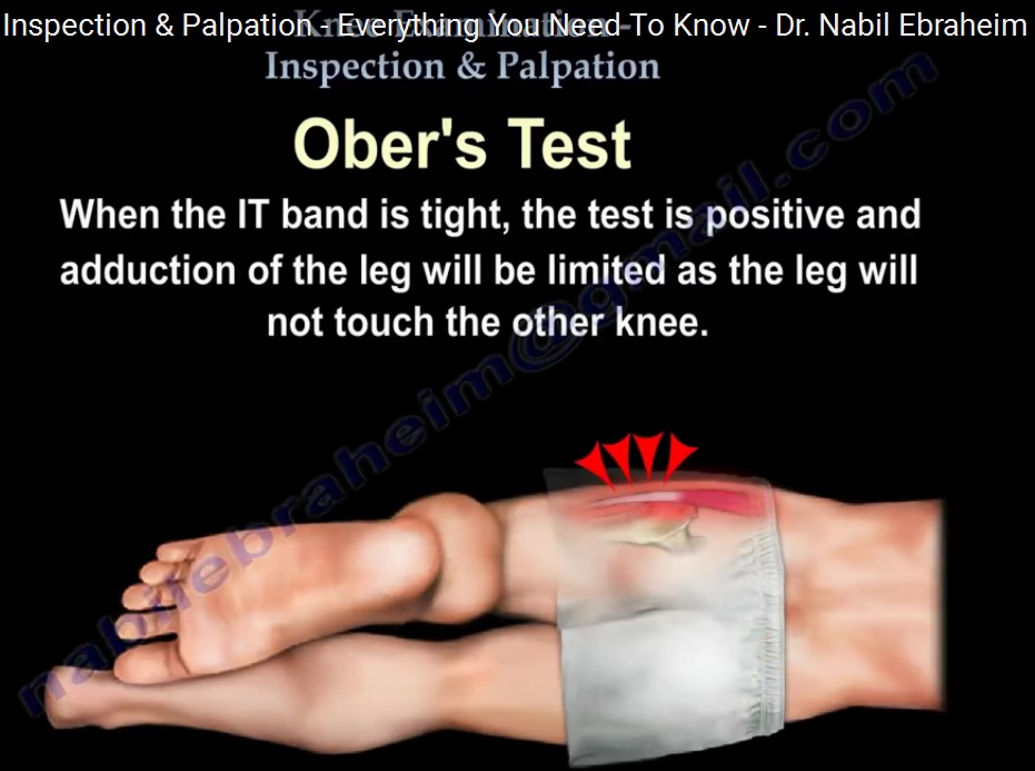
• OBERS TEST -ITB CONTRACTURE
• PHELPS -GRACILIS
• POPLITEAL ANGLE – HAMSTRING TIGHTNESS
• NOBLE TEST – ITB FRICTION SYNDROME
• PIPKIN SIGN SYNOVIAL PLICA
Examination of the Joint Above and Below
– Hip joint and ankle-foot complex
Examine opposite knee
Lymph Node Examination
– Inguinal and popliteal
Neurovascular assessment
Leave a Reply