Popular answer may not imply right answer. Answer will be published by the end of…
Comments
deyadeensays
answer c
CHANDRAKANTA NAYAKsays
ansawer is Pyknodysostosis . this is adiagnoses of exclusion. though pyknodysostosis commonly presents with repeated fractures following trivial trauma but cases reported in india didnt present with history of multiple fractures .generaly it presents with short stature ,generalized hypersostosis , hypoplastic distal phalanges , obtuse mandibular margin etc
adminsays
The combination of recurrent fractures and Osteosclerosis point towards a diagnosis of Ostepetrosis and Pyknodysostosis. Since Osteopetrosis is not there among the choices, the correct answer is Pyknodysostosis
HAMDOLLAH KIANFARDsays
sclerosis of medulla and non union of fx :the most probable dx is osteoporosis
Ganesh Singh Dharmshaktusays
The radiographic image resembles that of a patient of osteopetrosis, however not listed as an option here.
MAYUR RABHADIYAsays
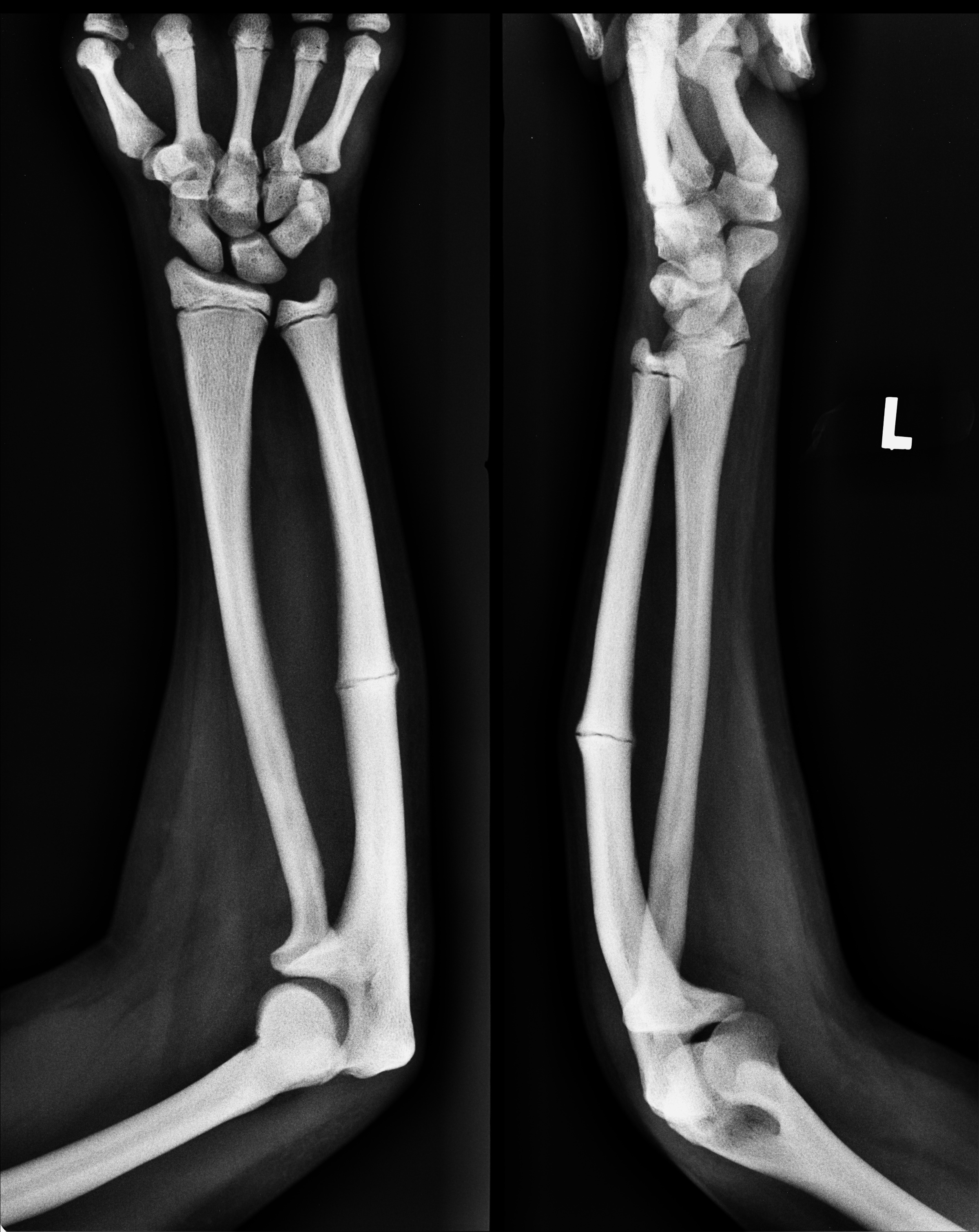
Xray of BB forearm with wrist and elbow joints AP and lateral of pt. With immature skeleton showing uniting fracture of middle third ulna with dense sclerosis of bones with v narrowed medullary canals and loss of cortico medullary differentiation. increased cortical thickness due to sclerosis. Condition most probably is pyknodysostosis.
Dr Chetan Naiksays
Plain xray AP/ lateral view left forearm in skeletally immature patient suggestive of loss of corticomedullary differentiation in both bone forearm with fracture midshaft ulna with hypertrophic fracture ends without any bridging callus formation- fracture line well distinguished
My most probable diagnosis is fracture midshaft ulna left forearm with delayed union/ hypertrophic non union most probable cause being osteopetrosis/ marble bone disease
Harsh jalansays
Answer complete undisplaced fracture of ulna with dorsal angulation…as there is history of multiple recurrent frcture so it can be d/t pyknodysostosis (by ruling out other options)
Kuldeep singhalsays
1. Plain x-ray forearm AP and Lat. View with wrist and elbow joint of a skeletally immature patient .
2. There is diffuse osteosclerosis , medullary canal is absent , thick cortex and a transverse fracture line seen in ulna mid shaft which shows signs of union .
3. Most probably a case of Osteopetrosis
D/D- skeletal flurosis
Ashish Upadhyaysays
Osteopokilosis as this growing child and sclerosis is present
Sachinsays
Pyknodysostosis
Ozairsays
Xray right forearm AP/lateral view of a skeletally immature patient showing an undisplaced ulna fracture with osteosclerosis with bridging callus on lateral aspect
D/D osteoporosis > pyknodysostosis
Dr Prabhat dubeysays
•Its plain X ray of
left forearm with wrist & elbow joint AP & lateral view
•Skeletally immature patient shows
• Generalized sclerosis of bone i.e chacky white in appearance
• loss of cortico- medullary differentiation in bone radius & ulna
• Complete undisplaced fracture of middle 3rd part of ulna bone
• contour of bone is maintained with no increase in soft tissue density
most problably mine radiological diagnosis is
– Pyknodiastosis ( d/t generalized osteosclerosis
D/D is
– metabolic disorder of bone
Osteopetrosis ( marble bone diseases)
Supreeth D Rsays
Pyknodysostosis
Ananta Narayan Pandasays
Plain x-ray ap and lat of left forearm showing both bones with wrist, elbow joint , carpals.some proximal part of metacarpals and distal third of humerus of skeletally immature patient showing fracture in midshaft of radius which is undisplaced with callus formation with hypertrophy at fracture ends in the background of diffuse sclerosis of both both bones of forearm with loss of corticomedullary differentiation.
Hypertrophy non Union or delayed union of fracture
With underground metabolic disease causing diffuse sclerosis
D/d
Osteopetrosis
Pyknodysosteosis
Hcsays
Pyknodysostosis
Sushruta Bhattacharyasays
Pyknodiostosis – characterised by brittle bones
Dr Sushruta Bhattacharyasays
This is a straight X-ray , showing AP and LatEral view of the lower end of humerus , radius and ulna , carpal bones , metacarpal bones and partly the proximal end of phalanges in a skeletaly immature person with a transverse fracture in mid shaft ulna without any displacement .
From the options – pyknodysostosis
Ashish Choprasays
It’s pyknodysostosis as suggested by diffuse sclerosis in radius & ulna ( long bones) & history of multiple fractures ( fractures occur in this condition with trivial trauma). Widespread sclerosis with maintenance of tabeculae as seen in metacarpals & carpal bones further confirm it .
Dr Prabhat dubeysays
Three differential for given x rays
i.e diffuse osteosclerotic lesion seen in children
As
pyknodiastosis AD disorder seen in children showing generalized osteosclerosis with pathologic # of long bone
For osteopetrosis
it also a AR in children .. show failure of function of osteoclast leading to
•generalized dense bone
• wideneing of metaphysis
•sandwich appearance vertebral bodies
Lastly fluorine toxicity
seen in all age groups
mainly exposed patient d/t drugs
show generalized osteosclerosis mainly in spine / pelvis
granular pattern
fragile bone prone for #
Suvratsays
I guess pyknodysostosis….By exclusion…As osteopetrosis not there…
Xray forearm AP and lateral of skeletally immature patient with sclerosis of metaphysis and diaphysis. Minimally displaced fracture shaft of ulna.
Sclerosis seen in hand bones including Metacarpals.
Pyknodysostosis a lysosomal storage disorder with sclerosed and brittle bones with frequent fractures.
Osteopetrosis
Its a bone disease that makes bones abnormally dense and prone to breakage (fracture). Researchers have described several major types of osteopetrosis, which are usually distinguished by their pattern of inheritance: autosomal dominant, autosomal recessive, or X-linked.
answer c
ansawer is Pyknodysostosis . this is adiagnoses of exclusion. though pyknodysostosis commonly presents with repeated fractures following trivial trauma but cases reported in india didnt present with history of multiple fractures .generaly it presents with short stature ,generalized hypersostosis , hypoplastic distal phalanges , obtuse mandibular margin etc
The combination of recurrent fractures and Osteosclerosis point towards a diagnosis of Ostepetrosis and Pyknodysostosis. Since Osteopetrosis is not there among the choices, the correct answer is Pyknodysostosis
sclerosis of medulla and non union of fx :the most probable dx is osteoporosis
The radiographic image resembles that of a patient of osteopetrosis, however not listed as an option here.
Xray of BB forearm with wrist and elbow joints AP and lateral of pt. With immature skeleton showing uniting fracture of middle third ulna with dense sclerosis of bones with v narrowed medullary canals and loss of cortico medullary differentiation. increased cortical thickness due to sclerosis. Condition most probably is pyknodysostosis.
Plain xray AP/ lateral view left forearm in skeletally immature patient suggestive of loss of corticomedullary differentiation in both bone forearm with fracture midshaft ulna with hypertrophic fracture ends without any bridging callus formation- fracture line well distinguished
My most probable diagnosis is fracture midshaft ulna left forearm with delayed union/ hypertrophic non union most probable cause being osteopetrosis/ marble bone disease
Answer complete undisplaced fracture of ulna with dorsal angulation…as there is history of multiple recurrent frcture so it can be d/t pyknodysostosis (by ruling out other options)
1. Plain x-ray forearm AP and Lat. View with wrist and elbow joint of a skeletally immature patient .
2. There is diffuse osteosclerosis , medullary canal is absent , thick cortex and a transverse fracture line seen in ulna mid shaft which shows signs of union .
3. Most probably a case of Osteopetrosis
D/D- skeletal flurosis
Osteopokilosis as this growing child and sclerosis is present
Pyknodysostosis
Xray right forearm AP/lateral view of a skeletally immature patient showing an undisplaced ulna fracture with osteosclerosis with bridging callus on lateral aspect
D/D osteoporosis > pyknodysostosis
•Its plain X ray of
left forearm with wrist & elbow joint AP & lateral view
•Skeletally immature patient shows
• Generalized sclerosis of bone i.e chacky white in appearance
• loss of cortico- medullary differentiation in bone radius & ulna
• Complete undisplaced fracture of middle 3rd part of ulna bone
• contour of bone is maintained with no increase in soft tissue density
most problably mine radiological diagnosis is
– Pyknodiastosis ( d/t generalized osteosclerosis
D/D is
– metabolic disorder of bone
Osteopetrosis ( marble bone diseases)
Pyknodysostosis
Plain x-ray ap and lat of left forearm showing both bones with wrist, elbow joint , carpals.some proximal part of metacarpals and distal third of humerus of skeletally immature patient showing fracture in midshaft of radius which is undisplaced with callus formation with hypertrophy at fracture ends in the background of diffuse sclerosis of both both bones of forearm with loss of corticomedullary differentiation.
Hypertrophy non Union or delayed union of fracture
With underground metabolic disease causing diffuse sclerosis
D/d
Osteopetrosis
Pyknodysosteosis
Pyknodysostosis
Pyknodiostosis – characterised by brittle bones
This is a straight X-ray , showing AP and LatEral view of the lower end of humerus , radius and ulna , carpal bones , metacarpal bones and partly the proximal end of phalanges in a skeletaly immature person with a transverse fracture in mid shaft ulna without any displacement .
From the options – pyknodysostosis
It’s pyknodysostosis as suggested by diffuse sclerosis in radius & ulna ( long bones) & history of multiple fractures ( fractures occur in this condition with trivial trauma). Widespread sclerosis with maintenance of tabeculae as seen in metacarpals & carpal bones further confirm it .
Three differential for given x rays
i.e diffuse osteosclerotic lesion seen in children
1) pyknodiastosis
2) osteopetrosis
3) flourine toxicity
As
pyknodiastosis AD disorder seen in children showing generalized osteosclerosis with pathologic # of long bone
For osteopetrosis
it also a AR in children .. show failure of function of osteoclast leading to
•generalized dense bone
• wideneing of metaphysis
•sandwich appearance vertebral bodies
Lastly fluorine toxicity
seen in all age groups
mainly exposed patient d/t drugs
show generalized osteosclerosis mainly in spine / pelvis
granular pattern
fragile bone prone for #
I guess pyknodysostosis….By exclusion…As osteopetrosis not there…
1.B prostrate
2.A OSTEOPOIKILOSIS
3.B spine
4.B pageta disease of bone
5.B.melorheostosis
6.C sarcoidosis
7.D Acro osteolysis
8.D osteopetrosis
9.A generalised osteopenia
10.C Healing NOF
Xray forearm AP and lateral of skeletally immature patient with sclerosis of metaphysis and diaphysis. Minimally displaced fracture shaft of ulna.
Sclerosis seen in hand bones including Metacarpals.
Pyknodysostosis a lysosomal storage disorder with sclerosed and brittle bones with frequent fractures.
Osteopetrosis
Its a bone disease that makes bones abnormally dense and prone to breakage (fracture). Researchers have described several major types of osteopetrosis, which are usually distinguished by their pattern of inheritance: autosomal dominant, autosomal recessive, or X-linked.