OrthopaedicPrinciples.com

Integrating Principles and Evidence

Integrating Principles and Evidence

  • Home
  • Editorial Board
  • Our Books
    • Evidence Based Orthopaedic Principles
  • Courses
  • Exams
  • Reviews
  • Live Program
  • Contact

Anatomy of the Meniscus

Courtesy: Prof Nabile Ebraheim, University of TOledo, Ohio, USA

ANATOMY OF MENISCUS

The meniscus is a cushion like structure made of cartilage which fits within the knee joint between the tibia and femur.
There are two menisci inside the knee joint:
1. Medial meniscus
2. Lateral meniscus

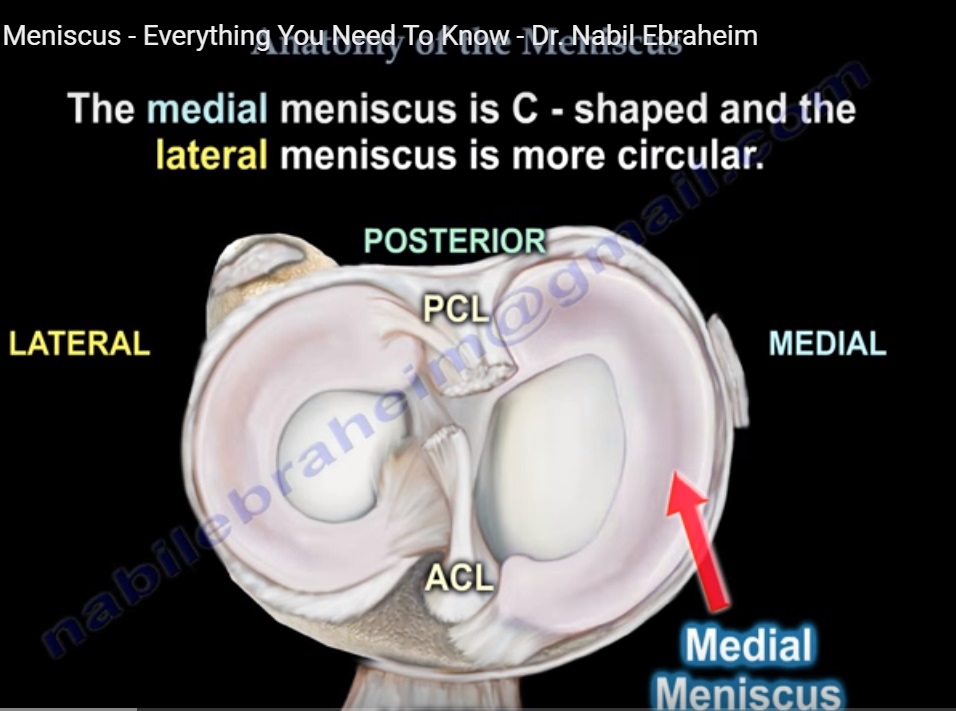
  • Medial meniscus is C shaped and the lateral meniscus is more circular .The medial meniscus covers 50% of the medial tibial plateau and the lateral meniscus covers 70% of the lateral tibial plateau.
  • With the medial meniscus, the anterior and posterior horns are separated from each other and from the anterior cruciate ligament. The posterior horn of the medial meniscus is the main secondary stabiliser of anterior translation.
  • The anterior and posterior horns of the lateral meniscus are closer to each other and near the insertion of the ACL .The transverse intermeniscal ligament connects the anterior horns of the lateral and medial menisci.
  • The meniscus is made up of type I collagen . The meniscus provides shock absorption and stability to the knee joint . The meniscus provides load sharing across the knee by increasing the contact area and decreasing the contact stress.
  • If the meniscus is removed the patient will develop arthritis of the knee joint .The contact stress increases 2-3 times when the meniscus is removed. The stress increases with the increased loss of meniscal tissue .
  • The meniscus helps to protect the knee joint, allowing the bones to slide freely on each other, however the meniscus limits flexion and extension extremes.
  • The meniscus transmits 50 degrees of force in extension and 90 degrees of force in flexion .Beyond 90 degrees , most of the force is transmitted to the posterior horn of the meniscus (flexion of the knee causes pain).

BLOOD SUPPLY

  • The blood supply of the meniscus decides the healing potential of the meniscus.
  • The peripheral 1/3 of the meniscus is vascular
  • It will heal if repaired
  • The inner 1/3 of the meniscus is not vascular and is nourished by synovial fluid
  • The middle third is red/white and it is avascular
  • The blood supply of the meniscus originates from the medial and lateral genicular arteries
  • The meniscus appears triangular in a cross section.

Traumatic tear of the meniscus may cause bleeding inside the knee joint . Swelling is usually gradual. It is different from an ACL tear that will have sudden , immediate swelling

NERVE SUPPLY

  • The peripheral portion of the meniscus has nerve supply.
  • It is well innervated in the posterior horn .(mechanoreceptors)
  • It provides proprioception during joint movement

APPLIED ANATOMY

  • Meniscal tears are commonly involving sports related activities with younger patients . Injury occurs from twisting or jumping activities such as with sports like football or skiing.
  • The tear usually is an acute , traumatic injury and it usually requires surgery.
  • In older individuals , tears are degenerative with a gradual onset of pain and the patient may not benefit from surgery.
  • Removal of the meniscus may lead to arthritis of the knee joint.
  • Tears of the medial meniscus occur three times more often than tears of the lateral meniscus .
  • The lateral meniscus is mobile and the medial meniscus is more fixed causing more tears to occur in the medial meniscus compared to the lateral meniscus.
  • The popliteus hiatus makes the lateral meniscus more mobile with less injuries.
  • The lateral meniscus is associated with a discoid meniscus and meniscal cyst. The lateral meniscus is also associated with acute injury to the anterior cruciate ligament(ACL).
  • The tear of the medial meniscus occurs more with degenerated tears and with ACL deficient knee. They are also associated with the baker’s cyst.

ANATOMICAL & SURGICAL CONSIDERATIONS
• The peroneal nerve is posterior to the biceps femoris tendon and must be protected during repair of the lateral meniscus.
• The saphenous nerve is posterior to the sartorius tendon and must be protected during repair of the medial meniscus

anatomy-of-meniscus

Post Views: 3,399

Related Posts

  • Sciatica Anatomy

    Courtesy: Prof Nabil Ebraheim University of Toledo, Ohio, USA Anatomy of sciatica The sciatic nerve…

  • Acetabular fractures: Applied Anatomy

    Courtesy: Saqib Rehman MD Director of Orthopaedic Trauma Temple University Philadelphia Pennsylvania USA

  • Anatomy of the Triceps

    Courtesy: Prof Nabil Ebraheim, University of Toledo, Ohio, USA

Leave a Reply Cancel reply

Your email address will not be published. Required fields are marked *

Follow Us

instagram slideshare

Categories

  • -Applied Anatomy
  • -Approaches
  • -Basic Sciences
  • -Cartilage & Meniscus
  • -Classifications
  • -Examination
  • -Foot and Ankle
  • -Foot and Ankle Trauma
  • -FRCS(Tr and Orth) tutorials
  • -Gait
  • -Hand and Wrist
  • -Hand and Wrist Trauma
  • -Hand Infections
  • -Hip and Knee
  • -Hip Preservation
  • -Infections
  • -Joint Reconstruction
  • -Knee Arthroplasty
  • -Knee Preservation
  • -Metabolic Disorders
  • -Oncology
  • -OrthoBiologics
  • -OrthoPlastic
  • -Paediatric Orthopaedics
  • -Paediatric Trauma
  • -Patellofemoral Joint
  • -Pelvis
  • -Peripheral Nerves
  • -Principles
  • -Principles of Surgery
  • -Radiology
  • -Rheumatology
  • -Shoulder and Elbow
  • -Shoulder and Elbow Arthroplasty
  • -Spine Deformity
  • -Spine Oncology
  • -Spine Trauma
  • -Spine, Pelvis & Neurology
  • -Sports Ankle and Foot
  • -Sports Elbow
  • -Sports Knee
  • -Sports Medicine
  • -Sports Medicine Hip
  • -Sports Shoulder
  • -Sports Wrist
  • -Statistics
  • -Technical Tip
  • -Technology in Orth
  • -Trauma
  • -Trauma (Upper Limb)
  • -Trauma Life Support
  • -Trauma Reconstruction
  • Book Shelf
  • Book Shelf Medical
  • Careers
  • Case Studies and Free Papers
  • DNB Ortho
  • Evidence Based Orthopaedic Principles
  • Evidence Based Orthopaedics
  • Exam Corner
  • Fellowships
  • Guest Editor
  • Guest Reviews
  • Image Quiz
  • Instructional Course Lectures
  • Journal Club
  • MCQs
  • Meetings and Courses
  • Multimedia
  • News and Blog
  • Plaster Techniques
  • Podcasts
  • Public Health
  • Rehabilitation
  • Research
  • Shorts and Reels

Book Shelf

Kendall’s Muscle Testing and Function 6th Edition

Kendall’s Muscle Testing and Function 6th Edition

By admin Leave a Comment

Get Book Kendall’s Muscles: Testing and Function, with Posture and Pain, 6th Edition, transforms this landmark Physical Therapy classic to prepare you for unparalleled clinical success in today’s practice. Timeless coverage of manual muscle testing, evaluation, and treatment meets the latest evidence-based practices, engaging imagery, and dynamic digital resources to create a powerful resource you […]

Popular Posts

  • Bone Screws in Orthopaedic Surgery
  • Silverskold Test
  • Piriformis Syndrome
  • Blood Supply of Long Bone
  • Movements of the Thumb

Recent Comments

  • RAJATABHA BISWAS on NUH Fellowship in Singapore
  • Runj on ESSKA Congress 2026
  • OT Hand Therapist on Interosseous Muscles Of The Hand
  • Badreddine on Rockwood and Green Fractures in Adults and Children- 10th Edition
  • Prof Dr P.sridhar MS Ortho,D Ortho on Rockwood and Green Fractures in Adults and Children- 10th Edition

Exam Corner

FRCS Orth Exam- Knee Arthroplasty

Courtesy: Zaid al Rab, FOunder, OrthoPass

MCQ Exam for the FRCS Orth 1

Courtesy: Zaid al Rub, Founder, OrthoPass

Postgraduate Entrance Exam Set 3

Get explanatory answers from our book

Postgraduate Entrance Exam Set 2

Get explanatory answers from our book

Main Menu

  • Orthopaedic Principles
  • Editorial Board
  • Orthopaedic Principles-A Review

Recent Posts

  • Anterior Tibial Tendon Tears
  • Endoscopic Lumbar Microdiscectomy
  • RAMP Lesion of the Knee
  • Osteochondritis Dissecans of the Knee
  • Dual Mobility Cups in Total Hip Replacement

Links

  • Join Our Editorial Board
  • Journals
  • Weblinks
  • Submit Your Conference
  • Disclaimer
Copyright@orthopaedicprinciples.com. All right rerserved.