OrthopaedicPrinciples.com

Integrating Principles and Evidence

Integrating Principles and Evidence

  • Home
  • Editorial Board
  • Our Books
    • Evidence Based Orthopaedic Principles
  • Courses
  • Exams
  • Reviews
  • Live Program
  • Contact

Thumb Metacarpal Base Fracture

Courtesy: Prof Nabile Ebraheim,University of Toledo, Ohio, USA

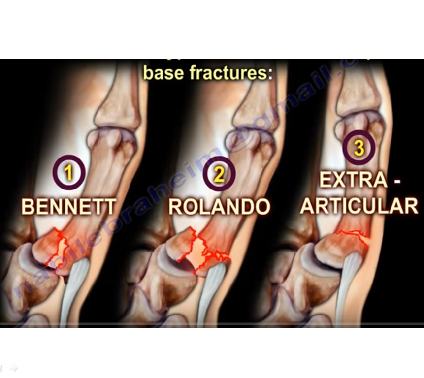
Fracture of the base of thumb metacarpal
The three types under it are the following:
1. Bennett fracture
2. Rolando’s fracture
3. Extra-articular fracture

Anatomy:

  • There are five metacarpal bones in the hand.
  • The first metacarpal bone is connected at the base to the trapezium at the first carpometacarpal joint.
  • The carpometacarpal joint is a saddle shaped joint.
  • The articulation between the first metacarpal bone and the trapezium gives inherent stability.
  • Trapezium has a concave articular surface and the first metacarpal has a convex articular surface.
  • Movement at the base of the thumb occurs around 2 axes:-
    1. Flexion and extension
    2. Abduction and adduction
    And rotation. Counting fingers with the thumb is called opposition.

Muscle insertions :
1. Abductor pollicis longus (inserted at the base of the first metacarpal)
2. Adductor pollicis (inserted at the base of the proximal phalanx)

Bennett fracture

  • It is a fracture dislocation at the base of the first metacarpal that extends into the carpometacarpal joint articulation.
  • There is volar-ulnar beak fracture fragment
  • Mechanism of injury:
    Axial overload along the 1st metacarpal with simultaneous flexion (as in the case of fist fight)

Deformity:
1. The distal part of the first metacarpal is adducted by the adductor pollicis muscle (ulnar nerve supply).
2. The metacarpal head is displaced into the palm.
3. The metacarpal shaft is displaced proximally by the abductor pollicis longus muscle (posterior interosseous nerve supply)
4. The metacarpal is therefore displaced dorsally, radially and supinated.
5. The small volar lip ulnar fragment remains in its position attached to the trapezium by the oblique ligament.
6. The main deforming forces are the abductor pollicis longus and adductor pollicis muscles.

Investigation:
X-ray plain or Robert view (hyperpronated AP thumb ) and true lateral view

Treatment:

  • Closed reduction percutaneous pinning or open reduction internal fixation if the fragment is large enough (more than 20%)
  • Reducing the 1st metacarpal to restore the articular surface: give longitudinal traction.
  • Pronate the metacarpal.
  • Then apply pressure over the thumb base to push it back to its base by abduction and extension of the metacarpal
  • The pinning will go from the thumb to the trapezium or from the thumb to the second metacarpal. Pinning may also go to both.
  • Apply thumb spica splint
  • Upto 2mm of articulation incongruity is accepted.

Note: The sensory branches of the radial nerve, radial artery and multiple tendons are in this area. Therefore the surgeon should be careful not to injure these structures during the surgery.

Reverse Bennett fracture

  • Intra-articular fracture of the base of the fifth metacarpal bone which extends into the carpometacarpal joint. The extensor carpi ulnaris has a pulling action on the 5th metacarpal.

Rolando fracture

  • Intra-articular, comminuted fracture through the base of the 1st metacarpal bone with same deforming forces as the Bennett fracture.
  • The base of the 1st metacarpal is split onto volar and dorsal fragments.
  • ‘Y’ fracture or transverse fracture
  • Sometimes three or more intra-articular fragments are present.

Treatment:

  • Difficult and controversial. Can be ORIF. Or closed reduction percutaneous pinning or external fixation.
  • Prognosis is worse than Bennett fracture and may end in arthritis.
  • Majority of cases do well even with residual displacement or incongruity.

Extra-articular fracture

  • It has the best outcome
  • Upto 30 degrees angulation is tolerated well. It can be managed with thumb spica splint
  • Angulation more than 30 degrees may lead to MCP hyperextension.
  • Therefore reduction and closed pinning is done. But in the case of high demand athletes, ORIF is done

Post Views: 7,731

Related Posts

  • Pilon Fracture

    Courtesy: International Foot and Ankle Course, Chennai

  • Knee Menisci

    Courtesy: Chris Beaulieu Stanford University, US

  • PIP Dorsal Fracture Dislocation

    Courtesy: Prof Nabile Ebraheim, University of Toledo, Ohio, United States

Comments

  1. Dr Priti Ashiwal says

    at

    Interesting article…thanks…

  2. Praveen sivakumar says

    at

    Very informative npractical thanq

  3. mahmoud awad says

    at

    many tnx

Leave a Reply Cancel reply

Your email address will not be published. Required fields are marked *

Follow Us

instagram slideshare

Categories

  • -Applied Anatomy
  • -Approaches
  • -Basic Sciences
  • -Cartilage & Meniscus
  • -Classifications
  • -Examination
  • -Foot and Ankle
  • -Foot and Ankle Trauma
  • -FRCS(Tr and Orth) tutorials
  • -Gait
  • -Hand and Wrist
  • -Hand and Wrist Trauma
  • -Hand Infections
  • -Hip and Knee
  • -Hip Preservation
  • -Infections
  • -Joint Reconstruction
  • -Knee Arthroplasty
  • -Knee Preservation
  • -Metabolic Disorders
  • -Oncology
  • -OrthoBiologics
  • -OrthoPlastic
  • -Paediatric Orthopaedics
  • -Paediatric Trauma
  • -Patellofemoral Joint
  • -Pelvis
  • -Peripheral Nerves
  • -Principles
  • -Principles of Surgery
  • -Radiology
  • -Rheumatology
  • -Shoulder and Elbow
  • -Shoulder and Elbow Arthroplasty
  • -Spine Deformity
  • -Spine Oncology
  • -Spine Trauma
  • -Spine, Pelvis & Neurology
  • -Sports Ankle and Foot
  • -Sports Elbow
  • -Sports Knee
  • -Sports Medicine
  • -Sports Medicine Hip
  • -Sports Shoulder
  • -Sports Wrist
  • -Statistics
  • -Technical Tip
  • -Technology in Orth
  • -Trauma
  • -Trauma (Upper Limb)
  • -Trauma Life Support
  • -Trauma Reconstruction
  • Book Shelf
  • Book Shelf Medical
  • Careers
  • Case Studies and Free Papers
  • DNB Ortho
  • Evidence Based Orthopaedic Principles
  • Evidence Based Orthopaedics
  • Exam Corner
  • Fellowships
  • Guest Editor
  • Guest Reviews
  • Image Quiz
  • Instructional Course Lectures
  • Journal Club
  • MCQs
  • Meetings and Courses
  • Multimedia
  • News and Blog
  • Plaster Techniques
  • Podcasts
  • Public Health
  • Rehabilitation
  • Research
  • Shorts and Reels

Book Shelf

Kendall’s Muscle Testing and Function 6th Edition

Kendall’s Muscle Testing and Function 6th Edition

By admin Leave a Comment

Get Book Kendall’s Muscles: Testing and Function, with Posture and Pain, 6th Edition, transforms this landmark Physical Therapy classic to prepare you for unparalleled clinical success in today’s practice. Timeless coverage of manual muscle testing, evaluation, and treatment meets the latest evidence-based practices, engaging imagery, and dynamic digital resources to create a powerful resource you […]

Popular Posts

  • Bone Screws in Orthopaedic Surgery
  • Silverskold Test
  • Piriformis Syndrome
  • Blood Supply of Long Bone
  • Movements of the Thumb

Recent Comments

  • RAJATABHA BISWAS on NUH Fellowship in Singapore
  • Runj on ESSKA Congress 2026
  • OT Hand Therapist on Interosseous Muscles Of The Hand
  • Badreddine on Rockwood and Green Fractures in Adults and Children- 10th Edition
  • Prof Dr P.sridhar MS Ortho,D Ortho on Rockwood and Green Fractures in Adults and Children- 10th Edition

Exam Corner

FRCS Orth Exam- Knee Arthroplasty

Courtesy: Zaid al Rab, FOunder, OrthoPass

MCQ Exam for the FRCS Orth 1

Courtesy: Zaid al Rub, Founder, OrthoPass

Postgraduate Entrance Exam Set 3

Get explanatory answers from our book

Postgraduate Entrance Exam Set 2

Get explanatory answers from our book

Main Menu

  • Orthopaedic Principles
  • Editorial Board
  • Orthopaedic Principles-A Review

Recent Posts

  • Anterior Tibial Tendon Tears
  • Endoscopic Lumbar Microdiscectomy
  • RAMP Lesion of the Knee
  • Osteochondritis Dissecans of the Knee
  • Dual Mobility Cups in Total Hip Replacement

Links

  • Join Our Editorial Board
  • Journals
  • Weblinks
  • Submit Your Conference
  • Disclaimer
Copyright@orthopaedicprinciples.com. All right rerserved.