Slipped Capital Femoral Epiphysis (SCFE)
Definition: Slipped capital femoral epiphysis refers to the atraumatic separation of the epiphysis in the epiphyseal plate of the femoral neck with displacement of the femoral head, usually in a medial and dorsal direction, during the pubertal growth spurt.
E. Müller was the first to describe this condition in 1888.
Aetiopathogenesis:
- The term slipped capital femoral epiphysis is a misnomer because the epiphysis is held in the acetabulum by the ligamentum teres, and thus it is actually the metaphysis that moves upward and outward while the epiphysis remains in the acetabulum.
- SCFE is associated with endocrine disorders, renal osteodystrophy, or with previous radiation therapy. But the most common variety is idiopathic SCFE
- Theories proposed for aetiology of Idiopathic SCFE are classified as Biomechanical and Biochemical
- Biomechanical factors: obesity, increased femoral retroversion, and increased physeal obliquity. These factors increase the shear stress across the physis
- Also, children with the disorder have deeper acetabula (increased CE angle of Wiberg), which causes increased stress across the physis (Kitadai, H. K et al..)
- Biochemical factors: association between endocrine dysfunctions like hypothyroidism, hypopituitarism, children who are receiving growth hormone supplementation, or who have hypogonadism
- Common in males: testosterone reduces physeal strength (oestrogen increases physeal strength)
- Age group affected: young adolescents with peak at 12- 13 years of age (range 9- 17 years)
- The left hip is affected twice as often as the right
- Histological abnormalities in SCFE: Chondrocyte clustering and disarray occur in a thickened hypertrophic zone. Defective collagen fibrils and defects in collagen banding also seen in this hypertrophic zone
- Proliferative Zone: changes in proteoglycan and glycoprotein concentrations, with increased glycoprotein staining in the territorial matrix and increased proteoglycan staining in the extraterritorial matrix
- SCFE occurs in the zone of hypertrophy
- Bilaterality: Incidence is around 18 to 50%. Higher in Black children compared to other races. Also higher incidence if one hip is treated with in-situ pinning (than with traditional casts)
- When bilateral slips occur, the second slip usually occurs within 12 to 18 months of the initial slip.
- Some authors believe that subtle SCFE that occur in childhood may be one cause of primary degenerative arthritis of the hip in adulthood.
- The direction of the slip is always posterior and often medial
Clinical Features:
- Children are often obese and present with pain in the hip (85%) or knee (15%)
- Paradoxical limp: when the patient bears weight on the involved limb, they will lean over that extremity during the stance phase of gait
- Patients with SCFE tend to keep their limb externally rotated while walking
- Knee Axilla sign: On attempted flexion of the hip, the patients leg goes into external rotation
- Internal rotation is lost. An attempted internal rotation will elicit pain
- Abduction is also restricted
- There is usually an exaggeration of extension (FFD is absent)
Type of Slips
1. Preslip: patient presents with weakness in the leg, limping, or pain in the groin or the knee on exertion. The most consistent positive finding is lack of internal rotation of the hip
2. Acute slip: presents with symptoms of less than three weeks and demonstration of an external rotation deformity, shortening, and marked limitation of motion secondary to pain. It is due to an abrupt displacement through the proximal physis in which there was a preexisting epiphysiolysis.
3. Chronic slip (most common- 90% cases)
4. Acute on chronic slip.
Loder Classification
- Stable: Patient is able to walk. Risk of osteonecrosis is 0%
- Unstable: Patient is unable to walk. Risk of osteonecrosis approaches upto 50%.
Triple deformity in SCFE
- Adduction due to coxa vara
- External rotation deformity
- Hyperextension
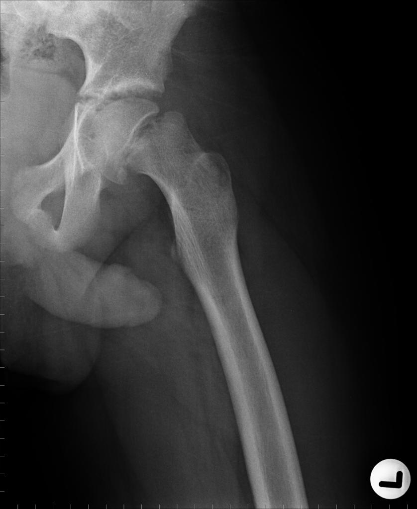
Radiology:
- The earliest sign is growth plate widening or lucency (epiphysiolysis).
- The earlier slip is the posterior slip and is best demonstrated in the lateral frog leg view.
1. Trethowan’s sign: A line (Klein’s line) drawn along the superior border of the femoral neck transects the femoral head. In SCFE the line passes above the femoral head.
2. Break in Shenton’s line
3. Capener’s sign: The posterior acetabular margin normally cuts the medial corner of the metaphysis. In slip the whole metaphysis remains lateral to the acetabular margin.
4. Metaphyseal blanch sign of Steel: A crescentic area of increased density in the metaphyses due to superimposition of displaced femoral head.
5. Articular portion of metaphysis will be excluded from the acetabulum.
6. Herndon’s hump: Bare area over the anterior and superior area of femoral neck that gets remodeled and becomes a hump.
7. Physeal neck angle: Normally is 87 degree. In slip it is reduced to 60 degree.
8. Head shaft angle(Southwick angle): Normal in AP view: 145 degree Lateral view: 170 degree.
MRI maybe helpful to diagnose a Preslip by showing a metaphyseal high signal next to the growth plate and growth plate widening.
Grading of Slip
• Mild slipping (grade I): Neck is displaced less than one-third of the diameter of the femoral head or when the head-shaft(Southwick angle) angle deviates from normal by 30° or less.
• Moderate slipping (grade II): The neck is displaced between one-third and one-half of the diameter of the femoral head, or the head-shaft angle deviates between 30 and 60 degrees from normal.
• Severe slipping (grade III): Characterised by neck displacement of more than half the diameter of the head, or deviation of the head-shaft angle of more than 60 degrees.
Treatment
Surgical treatment is the standard of choice.
Options
1. In Situ Pinning:
- Currently the most often used treatment for mild, moderate, and some severe acute or chronic SCFE.
- This is done by inserting one or more screws or pins across the growth plate, regardless of the severity of the slip.
- Advantages of single-screw fixation include percutaneous placement with minimal soft-tissue injury; a high success rate; a high patient satisfaction rate; and a low incidence of slip progression, osteonecrosis, and chondrolysis.
- A fracture table is used most often
- For ideal screw positioning, the bone is entered on the anterolateral aspect of the femoral neck in order to allow fixation perpendicular to the physis and to prevent hardware penetration through the posterior femoral neck
- A minimum of 4-5 screw threads should cross the fracture site
- The “approach-withdrawal phenomenon”, whereby the foot is rotated from maximum internal rotation to external rotation and observing the image intensifier helps to prevent screw penetration into the joint(9)
- The instant of change from approach(towards the joint) to withdrawal(away from joint) identifies the view in which the screw tip is shown in its true position
- Injection of arthrographic dye through the hardware under fluoroscopic control and bone endoscopy are two ways that have been reported for checking for pin penetration when high-quality radiographic images cannot be obtained intraoperatively(10,11)
2. Open epiphysiodesis
3. Proximal femoral osteotomy is indicated in high-grade slipped capital femoral epiphysis that does not remodel sufficiently with growth, despite treatment.
Femoral neck osteotomy (proximal/distal)
• Proximal neck osteotomy
Dunn, Fish and Martin Osteotomies
– Proximal neck osteotomies are associated with a higher rate of AVN
• Distal neck
– Kramer
– Craig
– Osteotomies are made distal to the major blood supply to the femoral head
4. Trochanteric osteotomy
- Campbell’s ball and socket osteotomy
- Tachdjian high subtrochanteric osteotomy
- Southwick biplanar intertrochanteric osteotomy
- Griffith geometric osteotomy.
– Trochanteric osteotomies are preferred to femoral neck osteotomies to correct alignment since it does not jeopardize the blood supply to the femoral head.
– Since the deformity is primarily in the sagittal plane a flexion intertrochanteric osteotomy is preferred.
– This minimizes risk to blood supply of the femoral head. It also helps to correct anterior impingement of neck or head-neck junction on the anterior acetabulum and rim.
– Anterior capsular release is also useful to release postoperative flexion deformity.
Complications
a) Chondrolysis: where degeneration of the hip articular cartilage occurs.
- Result from iatrogenic malposition (permanent penetration) of pins or screws used for fixation of slipped capital femoral epiphysis
- It may be painful and may progress to severe joint narrowing and degenerative changes within 6 months
- Cartilage is replaced by fibrous tissue, the joint capsule thickens and contracts, and joint motion is lost.
- Typically, the joint stiffens in flexion, abduction, and external rotation.
- X-rays: joint space narrowing, irregularity, and subchondral sclerosis and regional osteoporosis from disuse
- Treatment: NSAIDs, aggressive physical therapy and ROM exercises, and observation.
- Capsular release in resistant cases.
- Around 50% patients recover satisfactory with painless motion. The other half may require hip fusion for symptomatic relief
b) Avascular Necrosis:
- Some patients with partial head involvement regain a painless hip after 1–2 years of symptoms.
- Another group of patients with painless but abnormal ROM may be treatable by intertrochanteric osteotomy to reorient the arc of motion.
- Long-term pain following avascular necrosis is treated by hip fusion
Ref:
1. Dunn, D. M., and Angel, J. C.: Replacement of the femoral head by open operation in severe adolescent slipping of the upper femoral epiphysis. J. Bone and Joint Surg., 60-B(3): 394-403, 1978
2. Kitadai, H. K.; Milani, C.; Nery, C. A.; and Filho, J. L.: Wiberg’s center-edge angle in patients with slipped capital femoral epiphysis. J. Pediat. Orthop., 19: 97-105, 1999
3. Klein, A.; Joplin, R. J.; Reidy, J. A.; and Hanelin, J.: Slipped capital femoral epiphysis. Early diagnosis and treatment facilitated by “normal” roentgenograms. J. Bone and Joint Surg., 34-A: 233-239, Jan 1952
4. Loder, R. T.; Richards, B. S.; Shapiro, P. S.; Reznick, L. R.; and Aronson, D. D.: Acute slipped capital femoral epiphysis: the importance of physeal stability. J. Bone and Joint Surg., 75-A: 1134-1140, Aug 1993
5. Loder, R. T.: The demographics of slipped capital femoral epiphysis. An international multicenter study. Clin. Orthop., 322: 8-27, 1996
6. Loder, R. T, Slipped Capital Femoral Epiphysis, Instructional Course Lectures. J. Bone and Joint Surgery 82-A:1170, 2000
7. Southwick, W. O.: Osteotomy through the lesser trochanter for slipped capital femoral epiphysis. J. Bone and Joint Surg., 49-A: 807-835, July 1967
8. Steel, H. H.: The metaphyseal blanch sign of slipped capital femoral epiphysis. J. Bone and Joint Surg., 68-A: 920-922, July 1986
9. Moseley C: The “approach-withdraw phenomenon” in the pinning of slipped capital femoral epiphysis. Orthopaedic Transactions 1985; 9:497.
10. Bassett GS. Bone endoscopy: direct visual confirmation of cannulated screw placement in slipped capital femoral epiphysis. J Pediatr Orthop 1993;13:159-163.
11. Lehman WB, Grant A, Rose D, et al. A method of evaluating possible pin penetration in slipped capital femoral epiphysis using a cannulated internal fixation device. Clin Orthop 1984; 186:65-70
Common questions regarding this topic would be
1. What are the indications for contralateral pinning for SCFE?
2. Which is the preferred osteotomy for chronic slips and why?
3. How would you manage Avascular Necrosis after SCFE?How would you choose from vascularised fibular grafting, redirectional femoral osteotomy and hip fusion for AVN?
Please read: Lovell and Winter, Tachdjian.
when earliest can u remove the implant if chondrolysis occurs
few thoughts on planning for SCFE..
my earlier post wasnt uploaded..well in short what i wanted to say was how i plan a acute SCFE.. plz post all of ur comments …
No forceful or excessive or repeated manipulation / open reduction to achieve complete reduction in acute SCFE…i will put the patient in fracture table (DHS like) with neutral / internal rotation and pin it in the position i have then which might have some residual mal reduction…two pins mandatory if its an unstable slip and if i have enough reduction to pass them…contralateral prophylactic pinning to be counselled with parents in case of secondary slip …my arguments for doing so……
1) if the reduction doesn’t come once i have the patient (who will be in GA) in fracture table it usually means a a/c on c/c SCFE..so repeated , excessive forceful manipulations is not gonna give much benefit but also carries the risk of chondrolysis. so even if i have a mild to moderate severe residual slip i wont re- manipulate but pin in-situ
2) i haven’t seen any one open an acute slip..both benjmain joseph and KV Menon says they both wont open cuz they r not sure to achieve reduction without compromising vascularity
3) the residual coxa vara / retroversion in an in-situ pinning can be tackled later with corrective ostetomy if needed
Indications for contralateral pinning for SCFE
Prophylactic pinning is commonly performed in certain patient groups
1.Underlying endocrine disease because of their high rate of contralateral slip.
2.Previous pelvic radiation, which included the contralateral hip in the field
3.Children younger than 10 years at the time of presentation because of potential Limb length discrepancy following unilateral pinning and the high risk of bilateral involvement in such young children
it would be nice to see comments with references..Eg., standard books and journals…
Reference : Lovell and Winter’s pediatric Orthopaedics
thats good…
1) Lets start with classic Lowe’s article in 1961 43-B: 688-699. JBJS (Br); which was based on 100 cases of slipped upper femoral epiphysis treated at the Robert Jones and Agnes Hunt Orthopaedic Hospital, Oswestry, and the Prince of Wales Orthopaedic Hospital, Cardiff, in twenty-five years. In it he states that manual traction and reduction increases risk of AVN
2) BRIAN H. CASEY et al in ‘72 British JBJS(54-B: 607-614) observed 32 acute SCFEs among the 160 SCFEs and followed them up. What they noted was intra-operative traction and manipulation witn anatomic reduction showed maximum incidence of AVN..The method with least incidence of complications for them was skin traction with medial rotation boot for 3- 4 days and then pinning them in- situ (with some residual mal reduction)
3) Bishop et al series in CORR 135(93-96)’78 showed the increased risk of poor outcome in cases which had over reduction( valgus position) after manipulation under GA and reduction, they advice in-stu pinning even for severely displaced acute slips and later osteotomy after skeletal maturity
4) Martin Herman et al series in CORR, 322(77-85)’96, in their study of management of acute severe unstable slips strongly recommends against manipulation and repeated attempts to attain anatomical reduction . they postulate its almost like a double crush to the blood supply of femoral epiphysis for which if the first assault was the severe slip then the second assault will be the reduction attempts
5) In Long-Term Follow-up of Slipped Capital Femoral Epiphysis
BY BRIAN T. CARNEY, J Bone Joint Surg Am. 1991;73:667-674 which followed up 155 hips for 41 yrs, thirty-nine hips in which the slip had been manipulated under anesthesia &reduced, the mean Iowa hip-rating was 72 points. Osteonecrosis developed in twelve of these hips (31 per cent) and chondrolysis, in eleven (28 per cent). For the 116 hips that had not been reduced, the mean Iowa rating was 85 points and Osteonecrosis developed in seven hips (6 per cent) and treatment of these lesions. chondrolysis, in fourteen (12 per cent).
in short to summarize, my line of management in an acute sever unstable slip (or for that matter any acute slip even if its not serve or unstable) is to pin it in-situ within 24 hrs after positioning the patient in fracture table with some medial rotation and no excessive manipulation/ traction under G.A. if it cant be done within 24 hrs then put in skin traction with medial rotation boot and pin in- situ as soon as possible. i prefer two pins but one pin also is ok if the slip is not reduced enough to pass two pins
how do u guys go bt managing an acute slip when it presents to u?
what are the indications for inter trochanteric osteotomies