Courtesy: Prof Nabil Ebraheim, University of Toledo, Ohio, USA

ANATOMY OFTHE FEMORAL TRIANGLE
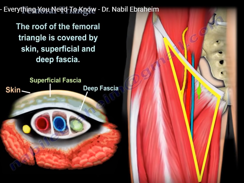
Femoral triangle is a superficial triangular space located on the anterior aspect of the thigh just inferior to the inguinal ligament .
The boundaries of the triangle include the medial border of the sartorius on the lateral aspect ,medial border of adductor longus muscle on the medial aspect and the base is formed by the inguinal ligament .The floor of the triangle is formed by iliacus muscle ,psoas major muscle ,pectineus muscle and adductor longus muscle and the roof is covered by skin ,superficial fascia and deep fascia .
The femoral triangle contains three important structures from lateral to medial which are femoral nerve, femoral artery and femoral vein .It also contains deep inguinal lymph nodes .The femoral nerve lies within the groove between the iliacus and psoas major muscles. Two other nerves which are also located within the femoral triangle are the lateral cutaneous nerve of the thigh and the femoral branch of the genitofemoral nerve .The lateral cutaneous nerve of the thigh crosses the lateral corner of the triangle and supplies the skin on the lateral part of the thigh .The femoral branch of the genitofemoral nerve runs in the lateral compartment of the femoral sheath and supplies majority of the skin over the femoral triangle.
In the anterior approach to the hip ,it is always safe to go lateral to the sartorius muscle in order to avoid the important structures within the femoral triangle.It is also important to avoid the lateral cutaneous nerve of the thighwhile performing this approach.
Mnemonic for arrangement of contents of Femoral Triangle from Lateral to Medial is “NAVI”
– Nerve Femoral
– Artery
– Vein
– Inguinal Lymph Node
Leave a Reply