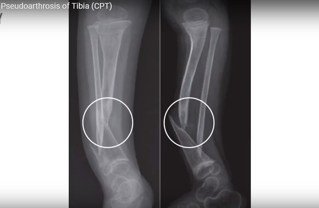
Courtesy: Sameer Qureshi MS, Consultant Orthopaedic Surgeon.
CONGENITAL PSEUDOARTHROSIS OF TIBIA
- Pseudarthrosis of the Tibia is a condition of unknown origin in which the discontinuity of the bone at the junction of middle and distal third is present at birth or developed thereafter during the growth period leading to abnormal mobility and creating an Illusion of a false joint
- 50% to 90% association of this disorder associated with neurofibromatosis
CLINICAL FEATURES
- Anterolateral or anterior angulation / bowing of leg at birth
- Foot deformity
- Ankle valgus
- Skin dimple
- Signs of neurofibromatosis : Café-au-lait spots
CLASSIFICATION
Multiple classification systems: Boyd, Crawford, Anderson
Boyd classification
Type I pseudarthrosis
- occurs with anterior bowing and a defect in the tibia present at birth.
Type II pseudarthrosis
- occurs with anterior bowing and an hourglass constriction of the tibia present at birth
- Spontaneous fracture, or fracture after minor trauma, commonly occurs before 2 years of age.
- The tibia is tapered, rounded, and sclerotic, and the medullary canal is obliterated.
- This type is the most common, is often associated with neurofibromatosis
Type III pseudarthrosis
- develops in a congenital cyst, usually near the junction of the middle and distal thirds of the tibia.
Type IV pseudarthrosis
- originates in a sclerotic segment of bone in the classic location without narrowing of the tibia.
- The medullary canal is partially or completely obliterated.
- An “insufficiency” or “stress” fracture develops in the cortex of the tibia and gradually extends through the sclerotic bone
Type V pseudarthrosis
- occurs with a dysplastic fibula.
- A pseudarthrosis of the fibula or tibia or both may develop.
Type VI pseudarthrosis
- occurs as an intraosseous neurofibroma or schwannoma that results in a pseudarthrosis.
- This is extremely rare.
Crawford classification
- Type I: the medullary canal is preserved and cortical thickening at the apex of the deformity might be observed; Good prognosis, Fracture may not occur in some patients
- Type II: presence of thinned medullary canal, cortical thickening, and trabeculation defect.
- Type III: cystic lesion, which may be fractured. Can experience early fracture and, therefore, require early treatment.
- Type IV: pseudarthrosis is present with tibial and possibly fibular nonunion
TREATMENT:
involves 3 principles
- resection of the entire pseudarthrosis and surrounding hamartomatous tissue,
- restoration of mechanical alignment
- intramedullary fixation.
These three basic principles often are augmented by a combination of
- primary shortening
- bone transport
- supplemental bone grafting
- bone morphogenetic protein
INTRAMEDULLARY FIXATION
- Most commonly used
- The pseudarthrosis is often quite distal in the tibia, making intramedullary fixation alone inadequate and unstable.
- Therefore, the ankle joint often must be crossed by the rod to provide
- The rod can migrate with growth, resulting in restoration of some ankle motion over time or can be surgically advanced to a position above the ankle once solid union has been achieved.
- For those lesions that appear more proximal in the tibia, it might be possible to avoid crossing the ankle joint.
- In these cases, larger rod diameter or an interlocking option could aid in stability.
VASCULARIZED GRAFT
- Resection of the pseudarthrosis with reconstruction using a free vascularized bone graft with either fibular or iliac crest grafts ..
ILIZAROV
- The Ilizarov approach with bone transport does offer the advantage of maintaining or gaining tibial length.
BONE MORPHOGENETIC PROTEIN
- Both currently available forms (rhBMP-2 and rhBMP-7) of this protein have been used.
- used in conjunction with other accepted forms of bony stabilization such as intramedullary fixation.
COMPLICATIONS
- STIFFNESS OF THE ANKLE AND HINDFOOT
- REFRACTURE
- VALGUS ANKLE DEFORMITY
- TIBIAL SHORTENING

Leave a Reply